Review
. 2008;3(4):637-58.
doi: 10.2147/copd.s4480.

Skeletal muscle dysfunction in patients with chronic obstructive pulmonary disease

Affiliations
Review

Skeletal muscle dysfunction in patients with chronic obstructive pulmonary disease

Ho Cheol Kim et al. Int J Chron Obstruct Pulmon Dis. 2008.

Abstract

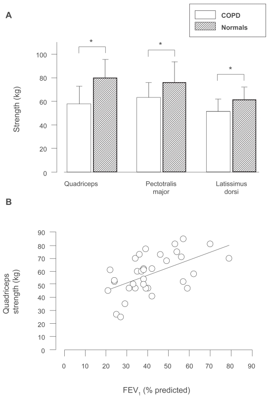
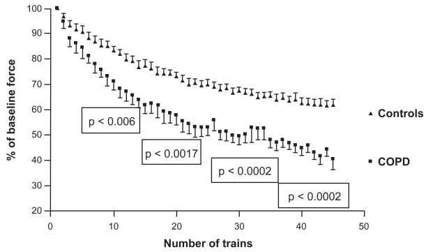
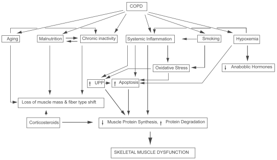
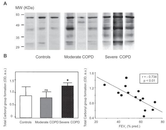
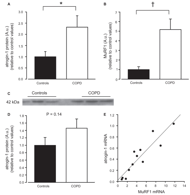
Chronic obstructive pulmonary disease (COPD) is a debilitating disease characterized by inflammation-induced airflow limitation and parenchymal destruction. In addition to pulmonary manifestations, patients with COPD develop systemic problems, including skeletal muscle and other organ-specific dysfunctions, nutritional abnormalities, weight loss, and adverse psychological responses. Patients with COPD often complain of dyspnea on exertion, reduced exercise capacity, and develop a progressive decline in lung function with increasing age. These symptoms have been attributed to increases in the work of breathing and in impairments in gas exchange that result from airflow limitation and dynamic hyperinflation. However, there is mounting evidence to suggest that skeletal muscle dysfunction, independent of lung function, contributes significantly to reduced exercise capacity and poor quality of life in these patients. Limb and ventilatory skeletal muscle dysfunction in COPD patients has been attributed to a myriad of factors, including the presence of low grade systemic inflammatory processes, nutritional depletion, corticosteroid medications, chronic inactivity, age, hypoxemia, smoking, oxidative and nitrosative stresses, protein degradation and changes in vascular density. This review briefly summarizes the contribution of these factors to overall skeletal muscle dysfunction in patients with COPD, with particular attention paid to the latest advances in the field.


Figures

Figure 1
Figure 1
A) Group mean values (±SD) for strength of quadriceps, pectoralis major and latissimus dorsi muscles obtained from normal subjects and COPD patients. Note significant reduction in strength in each of three muscle groups in patients with COPD, as compared to normal subjects. *P < 0.05. B ) Relationship between FEV1 (percentage of predicted) and quadriceps muscle strength showing significant positive relationship (r = 0.55, p < 0.0005) Copyright © 1998. Adapted from Bernard S, LeBlanc P, Whittom F, et al 1998. Peripheral muscle weakness in patients with chronic obstructive pulmonary disease. Am J Respir Crit Care Med, 158:629–34. Abbreviations: COPD, chronic obstructive pulmonary disease; FEV1, forced expiratory volume in one second.
Figure 2
Figure 2
Force decline of quadriceps femoris muscle during repetitive magnetic stimulation of femoral nerve in control subjects and COPD patients. The curves were significantly different at 10, 20, 30, and 40 trains Copyright © 2007. Adapted from Swallow EB, Gosker HR, Ward KA, et al 2007a. A novel technique for nonvolitional assessment of quadriceps muscle endurance in humans. J Appl Physiol, 103:739–46. Abbreviation: COPD, chronic obstructive pulmonary disease.
Figure 3
Figure 3
Proposed mechanisms of skeletal muscle dysfunction in COPD patients. Abbreviations: COPD, chronic obstructive pulmonary disease; UPP, ubiquitin/proteasomal pathway.
Figure 4
Figure 4
A) Representative examples of protein oxidation (total carbonyl groups) in diaphragms of control subjects and patients with moderate and severe COPD. B) Mean values ± SD of total carbonyl formation higher in the patients with severe COPD, compared to control subject muscles (*p = 0.05). No difference in total diaphragmatic carbonyl formation between patients with moderate COPD and control subjects (ns = nonsignificant). Among overall patients with COPD, optical densities of total carbonyl group formation significantly correlated with FEV1 (% predicted). Note that 14 COPD patients are depicted (two mild, six moderate, six severe) in correlation graph. Patients with moderate COPD have FEV1 and FVC of 62 ± 4 and 72% ± 11% of predicted, respectively. Patients with severe COPD have FEV1 and FVC of 42 ± 7 and 54% ± 9% of predicted, respectively. Copyright © 2005. Adapted from Barreiro E, de la Puente B, Minguella J, et al 2005a. Oxidative stress and respiratory muscle dysfunction in severe chronic obstructive pulmonary disease. Am J Respir Crit Care Med, 171:1116–24. Abbreviations: COPD, chronic obstructive pulmonary disease; FEV1, forced expiratory volume in one second; FVC, forced vital capacity.
Figure 5
Figure 5
A) Atrogin-1 mRNA expression; B) MuRF1 mRNA expression; C) representative Western blot for atrogin-1; D) atrogin-1 protein expression, all from quadriceps muscles of control subjects and COPD patients. Expression data relative to control values and presented in AU. mRNA data normalized for RPLPO, cytoplasmic protein contents normalized for α-tubulin. E) Significant correlation between atrogin-1 and MuRF1 mRNA expressions (r2 = 0.84; p < 0.001) in COPD patients. Values are mean ± SEM; *p < 0.05; +p < 0.001. Copyright © 2007. Adapted from Doucet M, Russell AP, Leger B, et al 2007. Muscle atrophy and hypertrophy signaling in patients with chronic obstructive pulmonary disease. Am J Respir Crit Care Med, 176:261–9. Abbreviations: AU, atomic units; COPD, chronic obstructive pulmonary disease; MuRF1, muscle-Specific RING finger protein 1; mRNA, messenger ribonucleic acid; RPLPO, large ribosomal protein; SEM, standard error of mean.
Figure 6
Figure 6
Top network of upregulated genes in quadriceps muscles of COPD patients, compared with control subjects. Figure created with Ingenuity Pathway Analysis System of microarray data from Debigare and colleagues (2008). Microarray data set used as an input file with RMA (robust multiarray analysis) threshold of 1.2 to specify upregulated genes. Filled nodes indicate upregulated genes in COPD patients. Empty nodes designate genes not detected by microarray data. Straight lines indicate direct relation while interrupted lines suggest indirect interactions. Abbreviations: AK1, adenylate kinase 1; ALDH2, aldehyde dehydrogenase 2 family, mitochondrial; COPD, chronic obstructive pulmonary disease; ERK, extracellular signal-regulated kinase; FHL2, four and half LIM domains 2; FHL3, cysteine and glycine-rich protein 3; FOXO1, forkhead box O1A (FKHR); FOXO3, forkhead box O3A (FKHRL1); FOX06, forkhead box 06; G0S2, putative lymphocyte G0/G1 switch gene; KLF9, Kruppel-like factor 9; NFκB, nuclear factor of kappa light polypeptide; P38MAPK, p38 mitogen-activated protein kinase; PDGF-BB, platelet-derived growth factor BB; SAA1, serum amyloid A1; SLC16A3, solute carrier family 16 (monocarboxylic acid transporters), member 3; T3-TR-RXR, T3-thyroid hormone receptor-retinoid X receptor complex; TMSB4X, thymosin beta 4, X-linked; TNNC1, slow troponin C; VIM, vimentin; UCP2, uncoupling protein 2, mitochondrial, proton carrier.

References

    1. Agusti A, Morla M, Sauleda J, et al. NF-kappaB activation and iNOS upregulation in skeletal muscle of patients with COPD and low body weight. Thorax. 2004;59:483–7. - PMC - PubMed
    1. Agusti AG, Sauleda J, Miralles C, et al. Skeletal muscle apoptosis and weight loss in chronic obstructive pulmonary disease. Am J Respir Crit Care Med. 2002;166:485–9. - PubMed
    1. Alexopoulou C, Mitrouska I, Arvanitis D, et al. Vascular-Specific growth factor mRNA levels in the human diaphragm. Respiration. 2005;72:636–41. - PubMed
    1. Allaire J, Maltais F, Doyon JF, et al. Peripheral muscle endurance and the oxidative profile of the quadriceps in patients with COPD. Thorax. 2004;59:673–8. - PMC - PubMed
    1. Allaire J, Maltais F, LeBlanc P, et al. Lipofuscin accumulation in the vastus lateralis muscle in patients with chronic obstructive pulmonary disease. Muscle Nerve. 2002;25:383–9. - PubMed

MeSH terms