Review
. 2009 Jun;46(6):811-20.
doi: 10.1016/j.yjmcc.2009.02.023. Epub 2009 Mar 9.

Morphological dynamics of mitochondria--a special emphasis on cardiac muscle cells

Affiliations
Review

Morphological dynamics of mitochondria--a special emphasis on cardiac muscle cells

Jennifer Hom et al. J Mol Cell Cardiol. 2009 Jun.

Abstract

Mitochondria play a critical role in cellular energy metabolism, Ca(2+) homeostasis, reactive oxygen species generation, apoptosis, aging, and development. Many recent publications have shown that a continuous balance of fusion and fission of these organelles is important in maintaining their proper function. Therefore, there is a steep correlation between the form and function of mitochondria. Many major proteins involved in mitochondrial fusion and fission have been identified in different cell types, including heart. However, the functional role of mitochondrial dynamics in the heart remains, for the most part, unexplored. In this review we will cover the recent field of mitochondrial dynamics and its physiological and pathological implications, with a particular emphasis on the experimental and theoretical basis of mitochondrial dynamics in the heart.


Figures

Figure 1
Figure 1. Model of the mechanism of mitochondrial fission and fusion in mammalian cells
Mitochondrial fission starts with DLP1 recruitment to the mitochondrial membrane. DLP1 can self-assemble in the cytosol. hFis1 and DLP1 are able to form a complex together, and it is thought that hFis1 serves as a transient receptor to recruit DLP1 to the mitochondria. After DLP1 is targeted to mitochondria, GTP-bound DLP1 forms a spiral completely around the mitochondrion. The constriction of the mitochondrial membrane may be driven by the assembly of DLP1 and/or a DLP1 conformational change driven by the hydrolysis of GTP into GDP + Pi. GTP hydrolysis allows the complete scission and disassembly of the DLP1 complex, thereby completing mitochondrial fission. Mitochondrial fusion requires opposing mitofusins to tether adjacent mitochondria together in a trans complex. GTP hydrolysis is essential for mitochondrial fusion. OPA1 is involved in inner mitochondrial membrane fusion and cristae remodeling. The mechanism by which the membranes come close enough for fusion, the actual fusion, or how mitofusins disassemble is not yet known.
Figure 2
Figure 2. Structure of proteins involved in mammalian mitochondrial fusion and fission
Abbreviations: GTP hydrolysis domain (GTPase), hydrophobic heptad repeats (HR), transmembrane segment (TM), dynamin-homology middle domain (Middle), GTPase effector domain (GED), tetratricopeptide repeats (TPR), proline-rich domain (PRD), really interesting new gene (RING). Mfn1/2 and OPA1 are involved in mitochondrial fusion. hFis1, DLP1, MARCH5, and MTP18 are involved in mitochondrial fission.
Figure 3
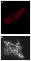
Figure 3. Mitochondrial morphology in adult and neonatal rat ventricular myocytes
A. One plane of mitochondria from an adult rat ventricular myocyte, visualized using TMRE under confocal microscope. Mitochondria within the cell are highly organized following patterns of the contractile apparatus. The mitochondria appear as uniform box-like shapes. B. Mitochondrial morphology of normal neonatal rat ventricular myocytes (Day 3 in culture) visualized by a mitochondrial-targeted red fluorescent protein (mRFP). There are two populations of mitochondria: thin filamentous or large globular. Unlike the adult myocytes, these mitochondria are disorganized and do not appear to be poised for excitation-contraction-metabolism coupling at this stage in development. The mitochondrial morphology may change in development to accommodate increased energy demands of the cell. C. Mitochondria from adult rat ventricular myocytes that are viewed from a longitudinal slice (left) appear as cylindrical or ovular shaped, whereas mitochondria that are viewed from a transverse slice (right are partially wrapped around myofilaments to different degrees.
Figure 4
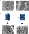
Figure 4. Different populations of mitochondria in the adult ventricular cardiomyocyte
A. Electron micrograph of adult rat ventricular myocyte. Abbreviations: S (Subsarcolemmal), I (Interfibrillar), N (Nuclear). Mitochondria directly under the sarcolemma vary in size, shape and organization. There is a mix of small and big mitochondria, with no distinct morphology and are not confined by contractile filaments. Interfibrillar mitochondria are constrained within contractile filaments, causing these mitochondria to be highly organized in a line with about one mitochondrion per sarcomere. These mitochondria tend to be big and ovular. Mitochondria around the nucleus tend to be highly disorganized and the size and shape vary greatly, ranging from small, spherical mitochondria to large globular mitochondria. B. Increased magnification of mitochondria viewed under the electron microscope. The filled arrow shows an example of soft membranes that can be found between adjacent cardiac mitochondria. The border between these mitochondria appears blurry and might indicate the early or intermediate stages of mitochondrial fusion or fission. The open arrow shows an example of defined membranes. Each mitochondrion has its own distinct outer membranes that are not in contact with other membranes. The borders and clearly defined with a visible space in between the outer membranes.
Figure 5
Figure 5. The role of mitochondria in excitation-contraction-metabolism coupling in the cardiac myocyte
During an action potential induced by pacemaker cells, L-type calcium channels open and allow Ca2+ to enter the cardiac myocyte. This Ca2+ influx activates ryanodine receptors on the SR to release calcium into the cytosol, causing a global increase in cytosolic Ca2+ concentration. This process is known as Ca2+-induced Ca2+ release. The cytosolic Ca2+ binds to Troponin C, which shifts the tropomyosin complex off of the actin binding site, exposing the site for the myosin head to bind to the actin filament. This is known as excitation-contraction coupling. Mitochondria are situated close to high Ca2+ microdomains. Ca2+ enters mitochondria down its electrochemical gradient due to the highly negative mitochondrial membrane potential that is maintained by the electron chain complexes. Ca2+ activates ATP production via Ca2+-activated dehydrogenases in the citric acid cycle and Ca2+-activated ATP synthase [136, 137]. ATP hydrolysis is needed for the myosin head to pull the actin filament to the center of the sarcomere. This unites metabolism to excitation-contraction coupling. For muscle relaxation to occur, intracellular Ca2+ is taken up by the SERCA pump or removed from the cell by the sodium-calcium exchanger or plasma membrane Ca2+ ATPase. This allows the tropomyosin complex to shift back over the active sites on the actin filaments.

References

    1. Scheffler IE. Mitochondria. 1. Wiley-Liss; 1999.
    1. Detmer SA, Chan DC. Functions and dysfunctions of mitochondrial dynamics. Nat Rev Mol Cell Biol. 2007 Nov;8(11):870–9. - PubMed
    1. Saotome M, Safiulina D, Szabadkai G, Das S, Fransson A, Aspenstrom P, et al. Bidirectional Ca2+-dependent control of mitochondrial dynamics by the Miro GTPase. Proc Natl Acad Sci U S A. 2008 Dec 19; - PMC - PubMed
    1. Suen DF, Norris KL, Youle RJ. Mitochondrial dynamics and apoptosis. Genes Dev. 2008 Jun 15;22(12):1577–90. - PMC - PubMed
    1. Szabadkai G, Simoni AM, Bianchi K, De Stefani D, Leo S, Wieckowski MR, et al. Mitochondrial dynamics and Ca2+ signaling. Biochim Biophys Acta. 2006 May–Jun;1763(5–6):442–9. - PubMed

Publication types