. 2009 May 22;284(21):14105-16.
doi: 10.1074/jbc.M807916200. Epub 2009 Mar 12.

Analysis of intracellular substrates and products of thimet oligopeptidase in human embryonic kidney 293 cells

Affiliations

Analysis of intracellular substrates and products of thimet oligopeptidase in human embryonic kidney 293 cells

Denise A Berti et al. J Biol Chem. .

Abstract

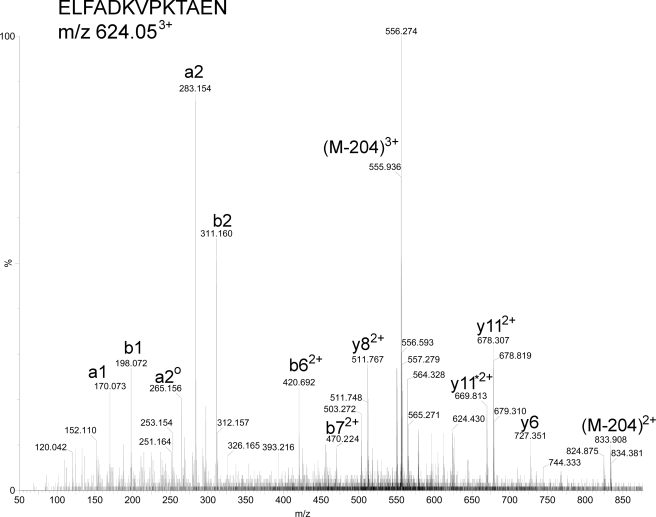
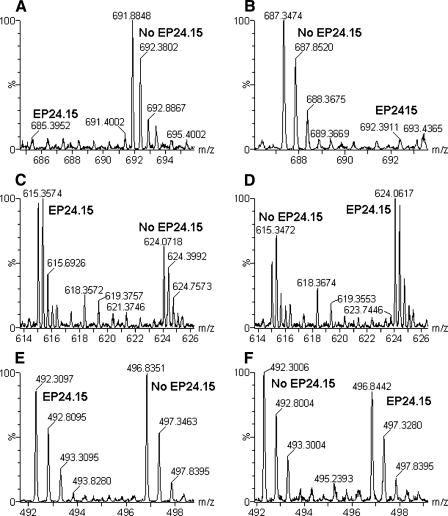
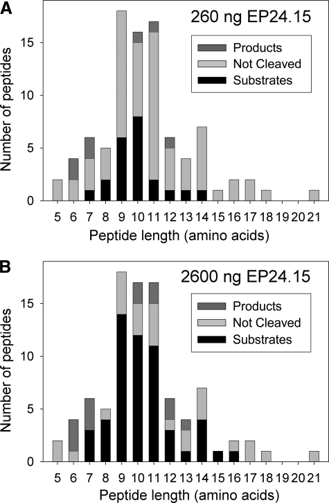
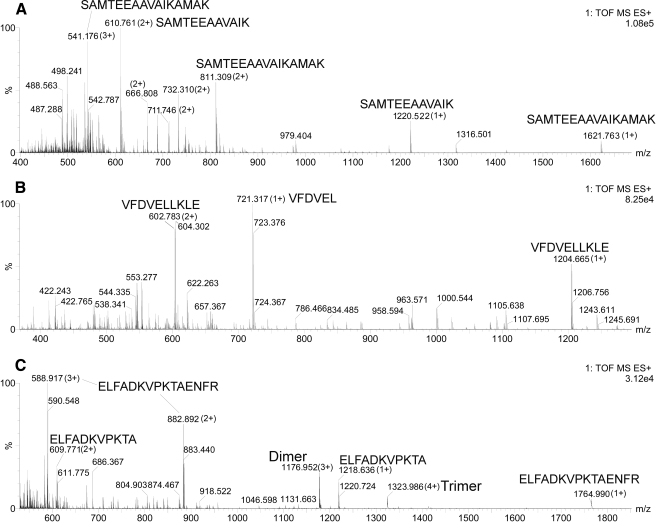
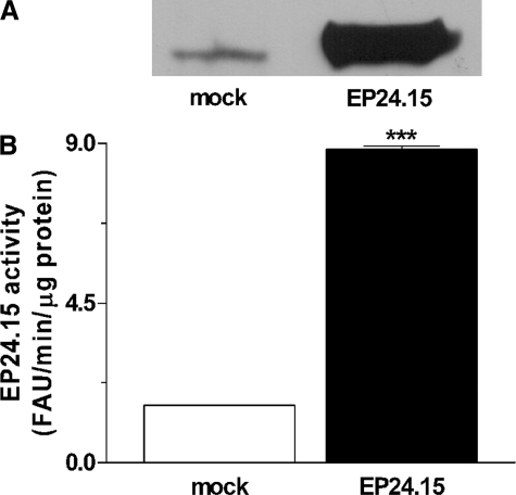
Thimet oligopeptidase (EC 3.4.24.15; EP24.15) is an intracellular enzyme that has been proposed to metabolize peptides within cells, thereby affecting antigen presentation and G protein-coupled receptor signal transduction. However, only a small number of intracellular substrates of EP24.15 have been reported previously. Here we have identified over 100 peptides in human embryonic kidney 293 (HEK293) cells that are derived from intracellular proteins; many but not all of these peptides are substrates or products of EP24.15. First, cellular peptides were extracted from HEK293 cells and incubated in vitro with purified EP24.15. Then the peptides were labeled with isotopic tags and analyzed by mass spectrometry to obtain quantitative data on the extent of cleavage. A related series of experiments tested the effect of overexpression of EP24.15 on the cellular levels of peptides in HEK293 cells. Finally, synthetic peptides that corresponded to 10 of the cellular peptides were incubated with purified EP24.15 in vitro, and the cleavage was monitored by high pressure liquid chromatography and mass spectrometry. Many of the EP24.15 substrates identified by these approaches are 9-11 amino acids in length, supporting the proposal that EP24.15 can function in the degradation of peptides that could be used for antigen presentation. However, EP24.15 also converts some peptides into products that are 8-10 amino acids, thus contributing to the formation of peptides for antigen presentation. In addition, the intracellular peptides described here are potential candidates to regulate protein interactions within cells.


Figures

FIGURE 1.
FIGURE 1.
Diagram of HEK293 peptide isotopic labeling. Experiment 1, HEK293 cellular peptide extract (160 μg) was incubated with active EP24.15 (260 or 2600 ng). As a control, the same amount of heat-inactivated EP24.15 was incubated for 20 min with 160 μg of HEK293 cellular peptide extract. After extract, but before purification of the peptides and subsequent analysis, the peptides were labeled with one of two isotopic forms of the TMAB label, either containing 9 atoms of deuterium (D9-TMAB) or only the hydrogenated form (D0-TMAB). The TMAB reagent labels the free amine group present on the N terminus of most peptides and also on the side chain of Lys residues (unless the amine group is acetylated or otherwise modified). For each concentration of enzyme, duplicate incubations were performed, and the duplicate digestion products were labeled with D0- or D9-TMAB in the opposite orientation from each other (runs 1 and 3 versus runs 2 and 4). Experiment 2, HEK293 cells were transfected with EP24.15-expressing cDNA or with the control cDNA plasmid. Extracts of EP2415-expressing and control cells were labeled with D0- or D9-TMAB, as indicated, for a total of four independent samples and LC/MS runs.
FIGURE 2.
FIGURE 2.
Representative peptide sequencing by ESI-MS/MS. This example shows the MS/MS analysis of the 3+ ion with an m/z of 624.05 that eluted from the reverse-phase column at 27.2 min and was identified as the peptidylprolyl isomerase A-derived peptide ELFADKVPKTAEN (monoisotopic mass of the unprotonated and untagged peptide = 1460.74 Da). Note that this peptide represents the D9-TMAB-labeled peptide shown in Fig. 3, C and D, and after collision-induced dissociation the a, b, and y fragments that contain TMAB tags have lost 68 Da (due to neutral loss of (C2H3)3N from the tag). In addition, the parent ion also shows neutral loss of 204 Da (= 3 × 68 Da) to produce the 3+ ion with m/z = 555.94 and the 2+ ion with m/z = 833.41. ao2 = a2 – H2O; y11* 2+= y2+11 – ammonia.
FIGURE 3.
FIGURE 3.
Representative MS spectra from H-TMAB and d-TMAB-labeled HEK293 cell extracts after incubation with purified EP24. 15. A, C, and E, data from Experiment 1, run 3; B, D and F, data from Experiment 1, run 4 (see Fig. 1). A and B, spectra of the peptide fragment from peptidylprolyl isomerase A subsequently identified by MS/MS as AVDGEPLGRVSF (supplemental Table 1). Note that this peptide decreases upon incubation with active EP24.15 and is therefore a substrate. C and D, spectra of another peptide fragment of peptidylprolyl isomerase A subsequently identified as ELFADKVPKTAEN (Fig. 2 and supplemental Table 1). Note that this peptide increases upon incubation with active EP24.15 and is therefore a product. E and F, spectra of the peptide subsequently identified as RLIVENL, a fragment of the protein splicing factor, arginine/serine-rich 4, 5, or 6 (supplemental Table 1). Note that this peptide is not affected by incubation with EP24.15 and is neither a substrate nor product. In all panels, EP24.15 represents the sample incubated with enzymatically active EP24.15, whereas no EP24.15 refers to the samples incubated with the heat-inactivated EP24.15.
FIGURE 4.
FIGURE 4.
Peptide length of EP24.15 substrates and products detected in the HEK293 cell peptidome after incubation with purified EP24.15. The number of peptides found in the HEK293 peptide extract is plotted versus peptide length. Peptides with an active/inactive enzyme ratio of less than 0.70 were considered substrates (i.e. levels decreased 30% or more with active enzyme). Peptides with an active/inactive enzyme ratio greater than 1.30 were considered products (i.e. levels increased 30% or more with active enzyme). Those with an active/inactive enzyme ratio from 0.70 to 1.30 were considered neither substrates nor products (not cleaved). A, when incubated with 260 ng of active enzyme, 22 peptides were substrates, 7 peptides were products, and 63 peptides were not cleaved. B, when the same HEK293 peptide extract was incubated with 2600 ng of active enzyme, 54 peptides were substrates, 13 peptides were products, and 26 peptides were not cleaved.
FIGURE 5.
FIGURE 5.
Analysis of cleavage of synthetic peptides by purified EP24.15. Peptides were incubated with purified EP24.15 and analyzed on LC/MS as described under “Experimental Procedures.” For each peptide, all MS spectra representing peptides were accumulated into a single spectrum as follows: A and C, the indicated spectrum represents all MS spectra from 20 to 30 min; B, indicated spectrum represents the sum of MS spectra from 25 to 31.5 min. A, most intense fragment from peptide SAMTEEAAVAIKAMAK was SAMTEEAAVAIK (1+ or 2+). B, most intense fragment from peptide VFDVELLKLE was VFDVEL (1+). C, most intense fragment from peptide ELFADKVPKTAENFR was ELFADKVPKTA (1+ or 2+).
FIGURE 6.
FIGURE 6.
Quantification of EP24.15 overexpression in HEK293 cells in Experiment 2. HEK293 cells were transiently transfected with empty pShooter (mock-transfected cells) or with pShooter coding for EP24.15 (EP24.15-transfected cells). A, proteins from crude cell extract (62.5 μg from mock cells and 5 μg from cells transfected with EP24.15) were separated by SDS-PAGE on an 8% polyacrylamide gel and transferred to nitrocellulose membranes, which were incubated with specific rabbit antiserum against EP24.15 (1:3000). After incubation with an anti-rabbit IgG-horseradish peroxidase-conjugated secondary antibody (1:3000), the immunoreactive bands were visualized by chemiluminescence. B, EP24.15 enzymatic activity was determined in triplicate using a continuous assay with a quenched fluorescent substrate as described under “Experimental Procedures.” EP24.15 activity was about six times greater in EP24.15 overexpressing cells then in the mock control group. ***, p < 0.001, statistically different from mock control group using Student's t test.

References

    1. Hershko, A., Ciechanover, A., and Varshavsky, A. (2000) Nat. Med. 6 1073–1081 - PubMed
    1. Ciechanover, A., and Schwartz, A. L. (2004) Biochim. Biophys. Acta 1695 3–17 - PubMed
    1. Lecker, S. H., and Goldberg, A. L. (2002) J. Physiol. (Lond.) 545 729. - PMC - PubMed
    1. Lippincott-Schwartz, J., Bonifacino, J. S., Yuan, L. C., and Klausner, R. D. (1988) Cell 54 209–220 - PubMed
    1. Geier, E., Pfeifer, G., Wilm, M., Lucchiari-Hartz, M., Baumeister, W., Eichmann, K., and Niedermann, G. (1999) Science 283 978–981 - PubMed

Publication types