Review
. 2009 Apr;9(4):259-70.
doi: 10.1038/nri2528.

Trophic macrophages in development and disease

Affiliations
Review

Trophic macrophages in development and disease

Jeffrey W Pollard. Nat Rev Immunol. 2009 Apr.

Abstract

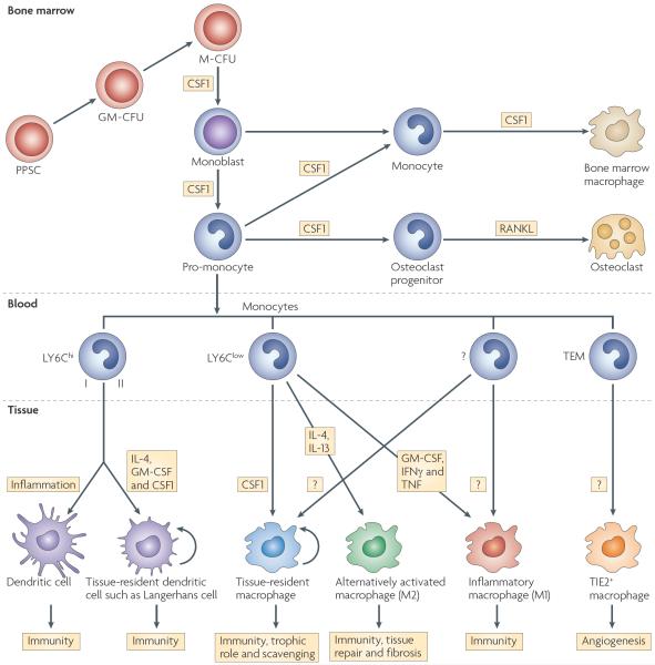
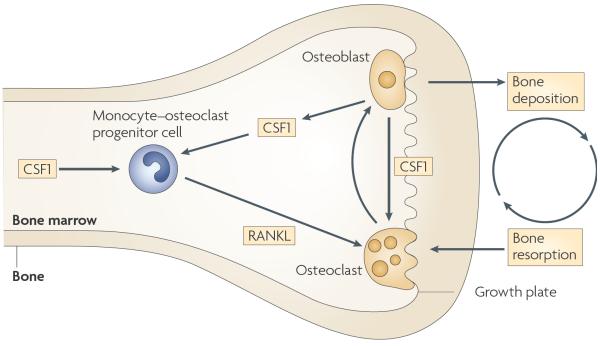
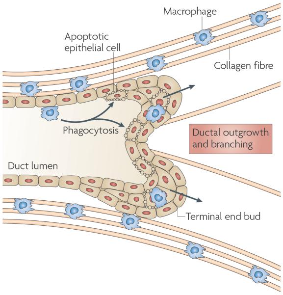
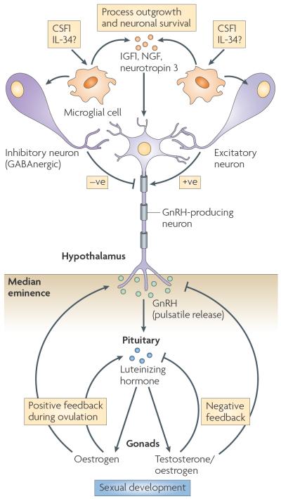
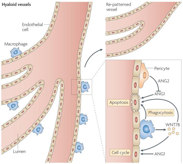
Specialized phagocytes are found in the most primitive multicellular organisms. Their roles in homeostasis and in distinguishing self from non-self have evolved with the complexity of organisms and their immune systems. Equally important, but often overlooked, are the roles of macrophages in tissue development. As discussed in this Review, these include functions in branching morphogenesis, neuronal patterning, angiogenesis, bone morphogenesis and the generation of adipose tissue. In each case, macrophage depletion impairs the formation of the tissue and compromises its function. I argue that in several diseases, the unrestrained acquisition of these developmental macrophage functions exacerbates pathology. For example, macrophages enhance tumour progression and metastasis by affecting tumour-cell migration and invasion, as well as angiogenesis.


Figures

Figure 1
Figure 1. The mononuclear phagocytic lineage and the control of its development by growth factors
Cells of the mononuclear phagocytic system (MPS) arise in the bone marrow, where they develop from pluripotent stem cells (PPSCs) through various multipotent progenitor stages: granulocyte/macrophage colony-forming unit (GM-CFU) to macrophage CFU (M-CFU) to monoblast to pro-monocyte. In the bone, osteoclast progenitors develop from these cells under the influence of colony-stimulating factor 1 (CSF1), and these differentiate in response to receptor activator of nuclear factor-κB ligand (RANKL) into osteoclasts. Another population differentiates into bone marrow macrophages also in response to CSF1, and the ex vivo culture of these cells and their progenitors is often used for macrophage studies. In addition, monocytes are released into the circulation. There is a growing body of evidence for an as yet undefined number of subpopulations of monocytes that have different developmental fates defined by the markers shown. LY6Chi monocytes consist of at least two types according to their expression of CXC-chemokine receptor 2 (CXCR2) and differentiate into dendritic cells of different types according to the state of inflammation and cytokine and/or growth factor exposure. Other as yet undefined types of monocytes are LY6Clow and differentiate into tissue-resident macrophages in response to CSF1; these have different names and functions according to their tissue residency (TABLE 1). Alternatively activated macrophages differentiate in response to parasitic infection, allergic conditions and during tissue repair through the effects of interleukin-13 (IL-13) and IL-4; these are also known as M2 macrophages. Inflammatory macrophages (also known as M1 macrophages) can also be found at sites of infection and injury, and these develop under the influence of GM-CSF, interferon-γ (IFNγ) and tumour necrosis factor (TNF). Immature macrophages can also differentiate into dendritic cells (not shown). In addition, TIE2-expressing monocytes (TEMs) give rise to TIE2+ macrophages that are involved in angiogenesis in tumours. It should also be noted that in addition to the growth factors shown here, many other ligands, particularly those that signal through Toll-like receptors, influence macrophage differentiation. In addition, the rigid lineage diagrams and growth factor assignments depicted here are for illustrative purposes only. In fact, there can be cross-differentiation of macrophage phenotypes during the evolution of an immune response, and tissue-restricted progenitors can expand within a tissue through proliferation. In addition, growth factors can act differently according to context and desired response, for example CSF1 receptor signalling is required for Langerhans-cell differentiation but CSF1 can act with IL-4 to inhibit dendritic-cell differentiation, at least in vitro. These complexities cannot be represented in such a simple lineage diagram but they are discussed in REFs 18,,.
Figure 2
Figure 2. The trophic role of macrophages in bone morphogenesis
Colony-stimulating factor 1 (CSF1) is required for the formation of a common monocyte–osteoclast progenitor cell in the bone marrow that then proliferates and differentiates in the presence of receptor activator of nuclear factor-κB ligand (RANKL) to form multinuclear osteoclasts. These remodel the bone that is laid down by osteoblasts. To coordinate these processes, osteoblasts produce CSF1, which not only mediates the correct positioning of osteoclasts but also affects their local function.
Figure 3
Figure 3. The trophic role of macrophages in ductal branching
In the mammary gland, macrophages are found in the stroma immediately adjacent to the growing terminal end bud (TEB) and are often associated with collagen fibres, which they help to form. Ablation of macrophages slows outgrowth and branching of the TEB into the fatty stroma, and this is associated with a disruption of collagen fibrillogenesis.
Figure 4
Figure 4. The trophic role of macrophages in neuronal patterning
Microglial cells are a specialized type of macrophage found in the brain. These cells respond to colony-stimulating factor 1 receptor (CSF1R) signalling to produce factors that are required for the establishment of neuronal connectivity. Depletion of CSF1 shows that, among other things, microglial cells regulate the hypothalamic–pituitary–gonadal axis through negative signalling from neurons that respond to γ-aminobutyric acid A (GABAA neurons; known as GABAnergic) and positive signalling from excitatory neurons, which allows gonadotrophin-releasing hormone (GnRH) to be released in a pulsatile manner into the median eminence. This induces the release of luteinizing hormone by the pituitary, which controls testosterone and oestrogen biosynthesis in the gonads. IGF1, insulin-like growth factor 1; IL-34, interleukin-34; NGF, nerve growth factor.
Figure 5
Figure 5. The trophic role of macrophages in angiogenesis and vascular remodelling
During eye development, the hyaloid vessel system regresses and is re-patterned. Macrophages closely associated with the vessels synthesize WNT7B, which stimulates vascular endothelial cells to enter the DNA synthesis phase of the cell cycle. In the presence of pericyte-produced angiopoietin 2 (ANG2), the survival signal from ANG1 is blocked and the endothelial cells undergo apoptosis and are phagocytosed by the macrophages. In this manner, vascular regression and proper patterning is achieved,.

References

    1. Tauber AI. Metchnikoff and the phagocytosis theory. Nature Rev. Mol. Cell Biol. 2003;4:897–901. - PubMed
    1. Kawai Y, Smedsrod B, Elvevold K, Wake K. Uptake of lithium carmine by sinusoidal endothelial and Kupffer cells of the rat liver: new insights into the classical vital staining and the reticulo-endothelial system. Cell Tissue Res. 1998;292:395–410. - PubMed
    1. van Furth R, et al. The mononuclear phagocyte system: a new classification of macrophages, monocytes, and their precursor cells. Bull. World Health Organ. 1972;46:845–852. - PMC - PubMed
    2. This article formalizes the concept of the MPS.

    1. Banchereau J, et al. Immunobiology of dendritic cells. Annu. Rev. Immunol. 2000;18:767–811. - PubMed
    1. Hume DA. Macrophages as APC and the dendritic cell myth. J. Immunol. 2008;181:5829–5835. - PubMed

Publication types