. 2009 Mar;5(3):e1000304.
doi: 10.1371/journal.pcbi.1000304. Epub 2009 Mar 13.

Detection of alpha-rod protein repeats using a neural network and application to huntingtin

Affiliations

Detection of alpha-rod protein repeats using a neural network and application to huntingtin

Gareth A Palidwor et al. PLoS Comput Biol. 2009 Mar.

Abstract

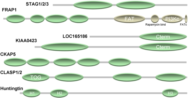
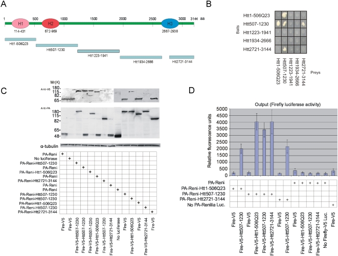
A growing number of solved protein structures display an elongated structural domain, denoted here as alpha-rod, composed of stacked pairs of anti-parallel alpha-helices. Alpha-rods are flexible and expose a large surface, which makes them suitable for protein interaction. Although most likely originating by tandem duplication of a two-helix unit, their detection using sequence similarity between repeats is poor. Here, we show that alpha-rod repeats can be detected using a neural network. The network detects more repeats than are identified by domain databases using multiple profiles, with a low level of false positives (<10%). We identify alpha-rod repeats in approximately 0.4% of proteins in eukaryotic genomes. We then investigate the results for all human proteins, identifying alpha-rod repeats for the first time in six protein families, including proteins STAG1-3, SERAC1, and PSMD1-2 & 5. We also characterize a short version of these repeats in eight protein families of Archaeal, Bacterial, and Fungal species. Finally, we demonstrate the utility of these predictions in directing experimental work to demarcate three alpha-rods in huntingtin, a protein mutated in Huntington's disease. Using yeast two hybrid analysis and an immunoprecipitation technique, we show that the huntingtin fragments containing alpha-rods associate with each other. This is the first definition of domains in huntingtin and the first validation of predicted interactions between fragments of huntingtin, which sets up directions toward functional characterization of this protein. An implementation of the repeat detection algorithm is available as a Web server with a simple graphical output: http://www.ogic.ca/projects/ard. This can be further visualized using BiasViz, a graphic tool for representation of multiple sequence alignments.


Conflict of interest statement

The authors have declared that no competing interests exist.

Figures

Figure 1
Figure 1. Detection of repeats in an alpha-rod protein.
Structure (alpha-backbone trace) of the 591 aa N-terminal fragment of human adaptor-related protein complex 2, beta 1 subunit, as forming part of the AP2 clathrin adaptor core (PDB code 2VGL chain B). Green and blue represent residues in alpha-helix and in disordered conformation, respectively. This structure has no residue in beta-strand conformation and is entirely composed of an alpha-rod of 14 repeats previously classified as HEAT repeats of type ADB . The label for each repeat indicates the following: repeat order, residue detected by the network, score of hit, and position relative to residue used for training. For example, “1 N24 0.84∶1” indicates that the residue detected for repeat #1 was N (amino acid code for asparagine) in position 24 of the sequence, with score 0.84, but that the residue in relative position 1 (that is, at 25) was the one used to train the network as being in the hinge. Ten out of the 14 repeats were detected, 8 of them with score> = 0.80. The inset shows repeats 12 (right, top) and 1 (right, bottom) with the residue used as positive in the training underscored. A coloured label indicates the residue identified by the network after training, which in both cases is not the one given in the training but others belonging to the hinge (E25 and S438). The figure was generated using NCBI's linked viewer, Cn3D .
Figure 2
Figure 2. Selected human protein families with alpha-rod repeats.
The cartoon summarizes the findings for seven human proteins. The green ellipses represent regions of alpha-rod repeats as deduced by a combination of our method, analysis of homologs, and iterative sequence analysis. Further details for each case, including an overview of repeat predictions and regions with amino acid bias overlaid to the multiple sequence alignment of the family using an update of the BiasViz software are available as supplementary Figure S3 in Text S1.
Figure 3
Figure 3. Study of interactions between fragments of huntingtin.
(A) Schematic overview of huntingtin fragments used in Y2H and LUMIER experiments. (B) The results obtained with the Y2H assays. (C) The expression of different fusion pairs was analyzed by Western blot using antibodies against V5-epitope (Invitrogen, 1∶5000, monoclonal antibody) and Protein-A (Sigma 1∶2000, polyclonal antibody); 15 µl from 100 µl of each cell extract was loaded onto SDS-PAGE gel. Detection with anti-tubulin antibodies was used as a loading control. (D) Firefly luciferase activities of immunopurified protein complexes in relative fluorescence units (RFU).
Figure 4
Figure 4. Hypothetical 3D structure of huntingtin.
The cartoon represents a hypothetical model of huntingtin interactions consistent with our results. (a) The N-terminus with the poly-Q tail (red arch) is followed by the H1 alpha-rod domain (residues 114 to 431, yellow cylinder), a small domain (432 to 671, blue), the H2 alpha-rod domain (672 to 969, yellow), a large domain (970 to 2666, green), the H3 alpha-rod domain (2667 to 2938), and a small C-terminal domain (2939–3144). (b) The three rods could assemble by coiling anti-parallel to each other with H2 in the middle: that would explain the interactions between H1 and H2, and between H2 and H3. (c) Formation of a huntingtin homodimer with a second molecule of huntingtin (gray) could happen through their H2 domains. The N-terminal poly-Q tail and the H1 domain remain exposed and can interact with other proteins, as previously reported . The figure was produced with Google SketchUp.

Similar articles

Cited by

References

    1. Andrade MA, Ponting CP, Gibson TJ, Bork P. Homology-based method for identification of protein repeats using statistical significance estimates. J Mol Biol. 2000;298:521–537. - PubMed
    1. Andrade MA, Perez-Iratxeta C, Ponting CP. Protein repeats: structures, functions, and evolution. J Struct Biol. 2001;134:117–131. - PubMed
    1. Andrade MA, Bork P. HEAT repeats in the Huntington's disease protein. Nat Genet. 1995;11:115–116. - PubMed
    1. Andrade MA, Petosa C, O'Donoghue SI, Muller CW, Bork P. Comparison of ARM and HEAT protein repeats. J Mol Biol. 2001;309:1–18. - PubMed
    1. Peifer M, Berg S, Reynolds AB. A repeating amino acid motif shared by proteins with diverse cellular roles. Cell. 1994;76:789–791. - PubMed

Publication types