. 2009;4(3):e4815.
doi: 10.1371/journal.pone.0004815. Epub 2009 Mar 13.

A GRFa2/Prop1/stem (GPS) cell niche in the pituitary

Affiliations

A GRFa2/Prop1/stem (GPS) cell niche in the pituitary

Montse Garcia-Lavandeira et al. PLoS One. 2009.

Abstract

Background: The adult endocrine pituitary is known to host several hormone-producing cells regulating major physiological processes during life. Some candidates to progenitor/stem cells have been proposed. However, not much is known about pituitary cell renewal throughout life and its homeostatic regulation during specific physiological changes, such as puberty or pregnancy, or in pathological conditions such as tumor development.

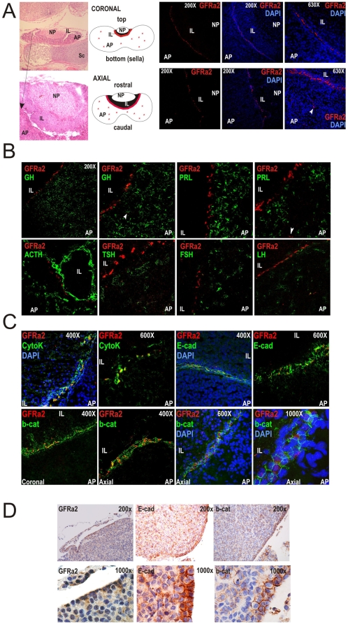
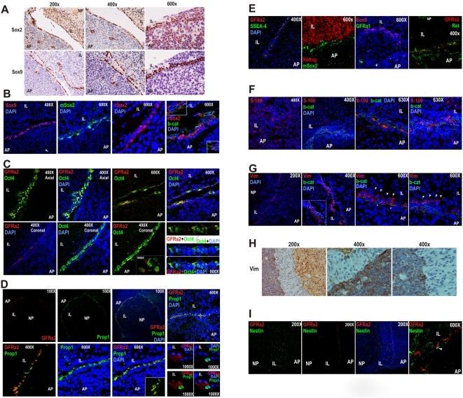
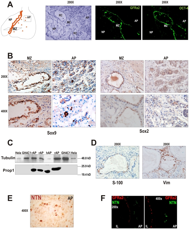
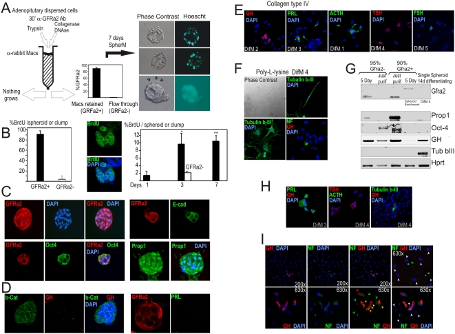
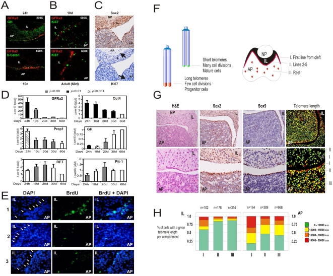
Principal findings: We have identified in rodents and humans a niche of non-endocrine cells characterized by the expression of GFRa2, a Ret co-receptor for Neurturin. These cells also express b-Catenin and E-cadherin in an oriented manner suggesting a planar polarity organization for the niche. In addition, cells in the niche uniquely express the pituitary-specific transcription factor Prop1, as well as known progenitor/stem markers such as Sox2, Sox9 and Oct4. Half of these GPS (GFRa2/Prop1/Stem) cells express S-100 whereas surrounding elongated cells in contact with GPS cells express Vimentin. GFRa2+-cells form non-endocrine spheroids in culture. These spheroids can be differentiated to hormone-producing cells or neurons outlining the neuroectoderm potential of these progenitors. In vivo, GPSs cells display slow proliferation after birth, retain BrdU label and show long telomeres in its nuclei, indicating progenitor/stem cell properties in vivo.

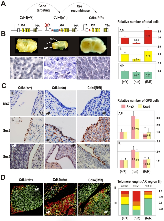
Significance: Our results suggest the presence in the adult pituitary of a specific niche of cells characterized by the expression of GFRa2, the pituitary-specific protein Prop1 and stem cell markers. These GPS cells are able to produce different hormone-producing and neuron-like cells and they may therefore contribute to postnatal pituitary homeostasis. Indeed, the relative abundance of GPS numbers is altered in Cdk4-deficient mice, a model of hypopituitarism induced by the lack of this cyclin-dependent kinase. Thus, GPS cells may display functional relevance in the physiological expansion of the pituitary gland throughout life as well as protection from pituitary disease.


Conflict of interest statement

Competing Interests: The authors have declared that no competing interests exist.

Figures

Figure 1
Figure 1. GFRa2+-expressing cells form a line of epithelial non-secretory cells in the adult pituitary of rats and mice.
A) Coronal and axial sections stained with Hematoxylin and Eosin (H&E) to show pituitary location under the hypothalamus and on top of the sphenoid sella turcica (Sc) and the disposition of the three pituitary lobes: adenopituitary (AP), intermediate lobe (IL) and neuropituitary (NP) where end-terminals of hypothalamic axons release ADH and Oxytocin. In the rat pituitary, GFRa2+ cells (red) arrange in a precise line in the frontier between the AP and the IL. Very few less intense cells are found dispersed through the AP (arrowhead). B) GFRa2+ cells [either lined or scattered (arrowhead)] do not express any pituitary hormone. C) GFRa2+ cells are epithelial cells with enhanced expression of Cytokeratins, E-cadherin and beta-Catenin. Coronal versus Axial sections demonstrates the orientation of the GFRa2 cells within the niche. In the coronal axis, GFRa2 or b-Catenin appear respectively as a thin line or a U-shaped green staining; in the axial axis GFRa2 appears as a broad surface while b-Catenin shows a ring shaped staining in a perpendicular orientation. D) Localization of GFRa2 cells and co-localization with E-cadherin and b-Catenin in mouse pituitaries.
Figure 2
Figure 2. The GPS Niche: GFRa2 cells express Prop1 and stem cell markers while neighbor cells express Vimentin.
A) Detection of Sox2 and Sox9 in the mouse and the rat pituitary (mSox2: mouse monoclonal and rSox2: rabbit polyclonal anti-Sox2 antibodies). (B). Sox2 signal co-localizes with b-Catenin. C) In rat pituitary, Oct4 is also expressed in the same line of cells, and co-localizes with GFRa2. D) Co-localization between GFRa2+ and Prop1 in the marginal zone between the AP and IL. Notice the nuclei positive for Prop1 surrounded by the GFRa2 membrane staining. E) GFRa2 cells co-localize with SSEA4, a glycolipid characteristic of Stem cells, but not with Nanog, which is restricted to the IL. GFRa2 cells do not express GFRa1 (which is however observed in somatotrophs) but weakly express the Ret receptor (Fig. S2). F) S-100 is expressed by the folliculostelate cells of the IL and AP of rat pituitary, but is also concentrated in around half of the b-Catenin/GFRa2 cells. G) Vimentin, a mesenchymal stem cell marker, is also expressed in the same niche as the GFRa2 cells but not in the same cells. Towards the IL, a parallel line of elongated cells (arrows) just beyond the b-Catenin/GFRa2 cells (asterisks) can be observed; fixation provokes sometimes the separation of both lines of cells (right panel). A similar Vimentin staining is seen in mouse pituitary (H). I) Although Nestin is expressed in the three portions of the pituitary, GFRa2 cells are negative for Nestin expression. Thin structures similar to axons apparently coming from the Nestin+ neuropituitary contact the GFRa2 cells (arrowhead).
Figure 3
Figure 3. The human marginal zone (MZ) of the pituitary contains a similar niche of GPS cells.
A) Cartoon representing the anatomy of the human pituitary with the anterior AP and a posterior NP; the boundary is called MZ and contains dilated structures usually called Rathke's remnant's Cysts (RC). Cells lining the RC express GFRa2 and Oct4. B) These cells also express Sox9 and Sox2. The human pituitary also contains small groups of Sox9+ or Sox2+ cells within the AP. C) Western blot detection of the pituitary specific factor Prop1 protein in rat (rAP) and human (hAP) pituitary, but not expressed in HeLa cells or a somatotroph pituitary cell line (GH4C1). D) S-100 is expressed in around half of the human GFRa2 cells lining the RC, similarly to what observed in the rat pituitary. Similarly, Vimentin+ elongated cells surrounded the GFRa2 epithelium (right panel). E) The GFRa2 ligand NTN is expressed in the human and rat (F) pituitary, and localizes exclusively at the AP.
Figure 4
Figure 4. Purified GFRa2+ cells form embryonic-like spheroids that differentiate into different ectodermic cell types.
A) Rat AP single-cell dispersions are prepared by treatment with Collagenase. The two fractions obtained, a GFRa2+ purified fraction (90% positivity for GFRa2 by immunofluorescence) and the Flow through GFRa2- (95% negativity for GFRa2), are then kept in SpherM. After 7 days, spheroids formed by small cells are observed in the GFRa2+ fraction. Some of they contain a hollow cavity while others are compact. A bunch of moving cilia is frequently observed in one pole of these spheroids (arrows; see videos in Supplementary Information). AP cell dispersion with trypsin does not result in viable spheroids as GFRa are extracellular receptors sensitive to trypsin treatment. B) Proliferation in the GFRa2+ and GFRa2- fractions after 5 days in the presence of BrdU. Center: A 7-day-old spheroid incubated with BrdU only for the last 12 hours before fixation. Right: BrdU uptake within growing spheroids incubated with BrdU during the last 12 hours before fixation. C) The spheroids are clonal (see Figure S3-A) and express GFRa2, Oct4, Prop1 or E-cadherin. D) These structures express b-Catenin but not hormones such as GH, PRL (and S3-B) or ACTH (data not shown). E–I) A single spheroid was transferred under the microscope to a collagen/poly-lysine-coated well and attached to the matrix with serum for 24 h. Spheroid structure disappear and cells spread through the well. Cells differentiate depending on the culture conditions into E) different pituitary secretory types (intermediate nuclei) or F) neurons (small or big nuclei) showing Tubulin beta III-+ cells+ or NF+ cells. G) mRNA expression of GFRa2/Prop1/Oct4 in the GFRa2+ purified fraction. The GFRa2+ fraction still have some contaminating secretory cells expressing GH. Five days later (spheroids) RNA expression of GFRa2 shifted (alternative splicing) while Prop/Oct4 were negative (even if the proteins were present). 14 days after differentiation of a single hand-picked spheroid, expression of either secretory (GH) or neuronal (Tubulin b-III) differentiation markers is detected. H) Double immunofluorescence in differentiated spheroids showed that differentiation is most frequently driven towards either secretory or neural phenotype. I) However, in some wells double GH/NF+ cells (orange arrows) together with single GH+ (red arrows) or NF+ (green arrows) or negative (white arrows) cells coexisted (Table 2).
Figure 5
Figure 5. GFRa2+ niche is present at birth and maintained through adulthood with reduced proliferation and long telomeres.
A) Newborn (24 h) and 10-days (10 d) old rat pituitaries present a GFRa2+/b-Catenin+ but GH- niche similar to that adult pituitaries (60 to 90-days old). B) 10 d pituitaries display abundant cell proliferation in the GPS niche, as seen by Ki67 staining, opposite to the adult rat (60 d) or mouse organs (C). D) Expression of GFRa2, Oct4, Prop1, GH, Ret and Pit1 in the AP of newborn and 10-, 20-, 30- and 60-day-old rats as detected by qRT-PCR. GPS progenitor markers (GFRa2, Oct4, Prop1) decrease with age while somatotroph markers (Ret, Pit-1) peak around puberty, day 10 to 20, or increase with grow to adulthood (GH). E) BrdU retaining in the GPS niche (arrows). Adult-pituitary nuclei within the niche retain BrdU injected in the rats as newborns. Three different animals (1–3) are depicted in the figure. F) Telomapping analysis of normal mouse pituitaries demonstrates a thin line of very long-telomere containing nuclei exactly in the first row of cells at the IL/AP boundary (regions I) matching the GPS niche. The following rows of cells towards the AP or the IL/NP present a shortening of the telomeres while the bulk of secretory cells have short telomeres characteristic of differentiated cells. G) Normal pituitaries were stained with H&E, Sox2 or Sox9 showing the GPS cells in the AP/IL boundaries (AP region I and IL region I) and some scattered groups through the AP (mostly in region III of the AP). Telomapping analysis as quantified in H) indicates that the region I of AP contains most long-telomere cells. This percentage progressively decreases in region II and III, where scattered GPS cells with long telomeres are found. In the IL, the only cells with long telomeres are also located in region I of the IL. AP, adenopituitary; IL, intermediate lobe; NP, neuropituitary.
Figure 6
Figure 6. Increased relative abundance of GPS progenitors and decreased formation of endocrine cells in Cdk4–null mice.
A) Alleles used in the analysis of GPS cells in a hypopituitarism model. The Cdk4-null allele Cdk4(n) is obtained by insertion of a neo-resistance cassette in Cdk4 intron 1. This mutation is rescued by expressing, through Cre recombination, a Cdk4R24C mutant allele that encodes a hyperactive Cdk4 . B) Cdk4-null mice display a hypoplastic pituitary much smaller than the wild type due to low cellularity and smaller size. These differences mostly affect the AP, containing around 5% cells of the wild-type pituitaries by 2-month age. This phenotype is rescued in the Cdk4(R/R) which displays a bigger pituitary with a 2-fold increase in the AP by 2-months. C) Genetic ablation of Cdk4 does not affect the structure of the stem cell niche. Moreover, the relative abundance of GPS cells is enlarged presenting more cell layers and 3–3.5-fold more GPS cells in the AP and 2-fold in the IL. D) The overall length of telomeres is significantly increased in the Cdk4-deficient AP distal region III. About 48% of these cells display long telomeres whereas this number is about 12% in wild-type or Cdk4R24C-rescued AP. AP, adenopituitary; IL, intermediate lobe; NP, neuropituitary.

Similar articles

Cited by

References

    1. Vankelecom H. Non-hormonal cell types in the pituitary candidating for stem cell. Semin Cell Dev Biol. 2007;18(4):559–570. - PubMed
    1. Allaerts W, Vankelecom H. History and perspectives of pituitary folliculo-stellate cell research. Eur J Endocrinol. 2005;153(1):1–12. - PubMed
    1. Horvath E, Kovacs K. Folliculo-stellate cells of the human pituitary: A type of adult stem cell? Ultrastruct Pathol. 2002;26(4):219–228. - PubMed
    1. Zhu X, Gleiberman AS, Rosenfeld MG. Molecular physiology of pituitary development: Signaling and transcriptional networks. Physiol Rev. 2007;87(3):933–963. - PubMed
    1. Vankelecom H. Stem cells in the postnatal pituitary? Neuroendocrinology. 2007;85(2):110–130. - PubMed

Publication types

MeSH terms

Substances