. 2009 Mar:Chapter 19:Unit 19.11.
doi: 10.1002/0471143030.cb1911s42.

Generation of transgenic mice

Affiliations

Generation of transgenic mice

Andrew Cho et al. Curr Protoc Cell Biol. 2009 Mar.

Abstract

This unit describes detailed step-by-step protocols, reagents, and equipment required for successful generation of transgenic mice using pronuclear injection. The experimental methods and practical tips given here will help guide beginners in understanding what is required and what to avoid in these standard protocols for efficiently generating transgenic mice.


Figures

Figure 19.11.1
Figure 19.11.1
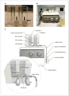
Purification of a transgenic construct using the sucrose gradient method. (A) Sucrose mixer with dimensions of 3-in. (W) × 2-in. (H) × 1 7/8-in. (D) is made by making two chambers of 5/8-in. (D) × 2-in. (H), which are connected together on the bottom with an outlet and two spigots that open and close controlling the sucrose flow into the ultracentrifuge tube. (B, C). A picture and schematic drawing showing how gradients are made from 10% to 40% sucrose.
Figure 19.11.2
Figure 19.11.2
Schematic diagram of the components necessary for preparing a mouth-controlled pipet device for handling zygotes. Rubber tubing with 1/8-in. i.d. and 1/32-in. wall is cut in three pieces, and the mouth piece, 0.2-μm syringe filter unit, 1-ml pipet cotton piece, and glass tubing are attached to each end of rubber tubing.
Figure 19.11.3
Figure 19.11.3
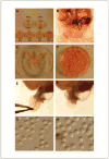
Sequence of embryo harvesting: (A) Two M16, three M2, and one M2 medium with hyaluronidase plates are prepared for handling or culturing embryos throughout the whole process of transgenic mice generation. (B, C) The reproductive organs: uterus, ovary, and oviduct are removed from the donor mice. (D) All the oviducts are collected in a single M2 dish. (E) The ampulla is located and torn apart with forceps under the microscope. (F) The zygotes and the cumulus cells are spilled into the M2 medium with hyaluronidase. (G) The zygotes are slowly separated from the surrounding cumulus cells. (H) Thoroughly washed zygotes are ready for microinjection.
Figure 19.11.4
Figure 19.11.4
Making of a holding pipet. (A) Microforge. (B) The schematic diagram of preparing holding pipets. The hand-pulled and diamond pencil-cut straight-edged pipet is selected and is brought closer to the heated glass ball to melt evenly to give a polished holding pipet with a small opening of ∼20 μm.
Figure 19.11.5
Figure 19.11.5
A schematic diagram for preparing injection needles. A glass tubing of 1.0-mm o.d. × 0.78-mm i.d. is inserted between two rubber pads and is pulled downward with the setting of 2.5 (downward force) × 16 (heat intensity) to prepare excellent and reproducible injection needles.
Figure 19.11.6
Figure 19.11.6
Microinjection setup. (A) A microinjection system. (B) Schematic diagram of the microinjection setup. A holding pipet is attached to the oil-pressured controller on the left side of the microscope whereas the injection pipet (needle) is attached to the automatic microinjector on the right side.
Figure 19.11.7
Figure 19.11.7
A schematic diagram depicting microinjection of transgenic DNA into the pronucleus. The size of the microinjected male pronucleus expands as the DNA is injected. A quick horizontal injection maneuver while minimizing damage to the zygote is critical for successful gene delivery.
Figure 19.11.8
Figure 19.11.8
A sequence of events for the microinjection technique. (A) Injection platform for microinjection. Note the rectangular M2 medium in the center that is overlaid with mineral oil. (B) A polished holding pipet is lowered onto the injection platform and a small volume of M2 medium is sucked into the pipet. (C) A zygote is held in place by the holding pipet and the injection needle is lowered and the tip is focused. (D) DNA solution is injected into the male pronucleus. Note the swelling of the pronucleus, which confirms the successful transfer of the transgene.
Figure 19.11.9
Figure 19.11.9
A schematic diagram of preparing transfer pipets for zygote transfers. A hand-pulled and diamond pencil-cut straight-edged pipet with ∼100-μm opening size is first loaded with the five alternate sequence of M16 medium and small bubbles and followed by fifteen to twenty tightly packed zygotes.
Figure 19.11.10
Figure 19.11.10
A sequence of events for zygote transfer into oviduct. (A) A surgical site is located and shaved. (B) A small skin incision is made and the ovary and fat pad are located before the muscle membrane incision is made. (C) The ovary and an oviduct are pulled out by the fat pad with the small metal clip. (D) The bursa is carefully peeled off and tucked under the ovary with a pair of forceps. (E) The space between the ovary and oviduct is opened with a pair of forceps while the tip of another forceps is inserted into the opening of the oviduct called the infundibulum for locating and testing prior to transferring zygotes with transfer pipet. (F, G) The transfer pipet is inserted into the infundibulum and the whole embryo content is blown slowly into the oviduct. (H) The muscle membrane is sutured and one drop of analgesia is administered to relieve pain during recovery. (I) the recipient mouse is placed on the slide warmer at 37°C.
Figure 19.11.11
Figure 19.11.11
A schematic diagram depicting zygote transfer into the infundibulum, which is often hidden within coils of oviduct. The infundibulum can be made visible by peeling off the bursa membrane that covers the ovary and by separating the space between the ovary and the oviduct using fine forceps.

References

Literature Cited

    1. Brinster RL, Chen HY, Trumbauer M, Senear AW, Warren R, Palmiter RD. Somatic expression of herpes thymidine kinase in mice following injection of a fusion gene into eggs. Cell. 1981;27:223–231. - PMC - PubMed
    1. Brown T. Southern Blotting. Curr Protoc Molec Biol. 1993;21:2.9.1–2.9.20.
    1. Conner DA. Transgenic mouse production by zygote injection. Curr Protoc Molec Biol. 2004;68:23.9.1–23.9.23. - PubMed
    1. Donovan J, Brown P. Parenteral injections. Curr Protoc Immunol. 2006a;73:1.6.1–1.6.10. - PubMed
    1. Donovan J, Brown P. Euthanasia. Curr Protoc Immunol. 2006b;73:1.8.1–1.8.4. - PubMed

Key References

    1. Brown GAJ, Corbin TJ. Transgenesis in the Mouse: Oocyte injection. In: Clarke AR, editor. Transgenesis Techniques. Methods in Molecular Biology. Vol. 180. Humana Press; Totowa, New Jersey: 2002. pp. 39–70. This chapter contains very useful information for the production of transgenic mice, especially for setting up a new transgenic facility. - PubMed
    1. Conner, 2004. See above.

      The most current information and methodologies for the generation of transgenic mice are found in this protocol.

    1. Nagy A, Gertsenstein M, Vintersten K, Behringer R. Manipulating the Mouse Embryo: A laboratory Manual. 3rd. Cold Spring Laboratory Press, Cold Spring Harbor; New York: 2003. A comprehensive guide for generating genetically altered mice along with excellent diagrams and illustrations.
    1. Overbeek PA. Factors Affecting Transgenic Animal Production. In: Pinkert CA, editor. Transgenic Animal Technology: A Laboratory Handbook. Academic Press; San Diego: 1994. pp. 72–109. A detailed description of different strains of mice, excellent husbandry practices, and transgenic phenomenology are discussed in this chapter.
    1. Pinkert CA. Introduction to Transgenic Animals. In: Pinkert CA, editor. Transgenic Animal Technology: A Laboratory Handbook. Academic Press; San Diego: 1994. pp. 3–11. A thorough and complete history of the development of transgenic technology, as well as a compendium for practical applications of transgenesis via pronuclear injections. - PubMed

LinkOut - more resources