. 2009 Mar 10:8:19.
doi: 10.1186/1476-4598-8-19.

delta-Catenin promotes prostate cancer cell growth and progression by altering cell cycle and survival gene profiles

Affiliations

delta-Catenin promotes prostate cancer cell growth and progression by altering cell cycle and survival gene profiles

Yan Zeng et al. Mol Cancer. .

Abstract

Background: delta-Catenin is a unique member of beta-catenin/armadillo domain superfamily proteins and its primary expression is restricted to the brain. However, delta-catenin is upregulated in human prostatic adenocarcinomas, although the effects of delta-catenin overexpression in prostate cancer are unclear. We hypothesized that delta-catenin plays a direct role in prostate cancer progression by altering gene profiles of cell cycle regulation and cell survival.

Results: We employed gene transfection and small interfering RNA to demonstrate that increased delta-catenin expression promoted, whereas its knockdown suppressed prostate cancer cell viability. delta-Catenin promoted prostate cancer cell colony formation in soft agar as well as tumor xenograft growth in nude mice. Deletion of either the amino-terminal or carboxyl-terminal sequences outside the armadillo domains abolished the tumor promoting effects of delta-catenin. Quantitative RT2 Profiler PCR Arrays demonstrated gene alterations involved in cell cycle and survival regulation. delta-Catenin overexpression upregulated cyclin D1 and cdc34, increased phosphorylated histone-H3, and promoted the entry of mitosis. In addition, delta-catenin overexpression resulted in increased expression of cell survival genes Bcl-2 and survivin while reducing the cell cycle inhibitor p21Cip1.

Conclusion: Taken together, our studies suggest that at least one consequence of an increased expression of delta-catenin in human prostate cancer is the alteration of cell cycle and survival gene profiles, thereby promoting tumor progression.


Figures
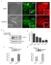
Figure 1
Figure 1
δ-Catenin expression is important for viable prostate cancer cell growth. A. Establishment of stable CWR22Rv-1 cells overexpressing δ-catenin and its effects on epithelial cell morphology. CWR22Rv-1 cells, showing epithelial morphology (a) when transfected with pEGFP as vector control (b), display expression of E-cadherin at the cell-cell junction (c). δ-Catenin overexpressing CWR22Rv-1 cells (e) interfere with the epithelial monolayer (d) and disrupt E-cadherin expression at the cell-cell junction (f). Bar, 30 μm. Inserts: selective higher magnification images for (b), (c), (e) and (f), respectively. Bar, 25 μm. B. δ-Catenin overexpression promotes, while its knockdown suppresses prostate cancer CWR22Rv-1 cell growth. a. Western blot analysis shows that the increased expression of δ-catenin in cells transfected with δ-catenin cDNA and reduced expression of δ-catenin in cells transfected with δ-catenin shRNA. Anti-actin staining is used as a loading control, and the molecular weight markers (kDa) are on the left. b. δ-Catenin shRNA transfection reduces viable cell numbers while δ-catenin overexpression by δ-catenin cDNA transfection increases viable cell numbers. Vector 1 and 2: pRS-GFP and pEGFP, respectively. shRNA 1 and shRNA 2: shRNA against δ-catenin sequences 1 and 2, * P < 0.05. c and d. δ-Catenin overexpression promotes cancer cell growth in PC-3 (c) and NCI-H1299 (d) cells. Inserts: Western blots showing PC-3 (c) and NCI-H1299 (d) cells with (+) or without (-) stable δ-catenin overexpression. Anti-actin staining is used as a loading control, and the molecular weight markers (kDa) are on the left.
Figure 2
Figure 2
δ-Catenin expression mediated anchorage-independent prostate cancer cell colony formation and tumor xenograft growth replies on the NH2- and COOH-terminal sequences outside armadillo domains. A. Soft agar assays showing that full length δ-catenin, but not its NH2- or COOH-terminal truncation mutants, promotes CWR22Rv-1 cell colony formation in vitro. Stable cells expressing vector control, full-length δ-catenin, ΔN280 and ΔC207 were plated in soft agar. Colonies were counted under the phase contrast light microscope in 1, 2, 3, and 4 weeks after they were plated. Results were derived from five independent experiments, each in duplicate. * P < 0.05. B. δ-Catenin overexpression promotes CWR22Rv-1 cells to grow tumors in nude mice. Shown here is a representative pair of tumor bearing mice expressing control vector and δ-catenin, respectively (n = 19). C. Full-length δ-catenin, but not its NH2- or COOH-terminal truncation mutants, promotes tumor xenograft growth in nude mice. Male athymic nude mice were inoculated subcutaneously with either the vector, full-length δ-catenin, ΔC207 or ΔN280 and were then allowed to grow for 5 weeks. Each week, the tumor volumes were measured and compared to each other. * P < 0.05. D. Fluorescent light microscopy showing the tumor cell morphology (a and c) and GFP positive CWR22Rv-1 cells expressing vector alone (b) and overexpressing δ-catenin (d). Bar, 50 μm.
Figure 3
Figure 3
A. Establishment of stable CWR22Rv-1 cells overexpressing full-length, ΔN280 and ΔC207 δ-catenin cDNAs. Top panel, schematic illustration of full-length, ΔN280 and ΔC207 δ-catenin cDNAs. Bottom panel, western blots showing the stable expression of full-length, ΔN280 and ΔC207 δ-catenin protein in CWR22Rv-1 cells. Molecular weight markers are shown on the left. B. GFP fluorescent images of CWR22Rv-1 cells stably expressing vector (a), fill-length δ-catenin (b, arrow points to cell-cell junction), ΔN280 (c, arrow points to cell-cell junction) and ΔC207 (d, arrow points to cytoplasmic distribution). Bar, 50 μm.
Figure 4
Figure 4
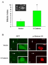
δ-Catenin expression promotes the entry of mitosis. A. Mitotic index of cells expressing vector alone as a control and cells overexpressing δ-catenin. Mitotic index is given as the percentage of cells entering mitosis determined by Hoechst staining. Insert: mitotic cells (arrows) showing strong Hoechst staining. B. Immunofluorescent light microscopy showing increased mitotic activity in δ-catenin expressing cells (c and d) in comparison to control vector transfected cells (a and b). Mitotic activity is determined as the number of GFP positive, transfected cells (a and c) intensely reactive with anti-phosphorylated histone H3 (p-Histone H3, b and d). Arrows: examples of GFP positive, transfected cells immunoreactive for phosphorylated histone H3. Bar, 100 μm; Bar in the insert, 5 μm.
Figure 5
Figure 5
Changes in gene profiles of CWR22Rv-1 cells expressing δ-catenin when compared to that of cells expressing vector alone. A. RT2 Profiler™ PCR Array. CWR22Rv-1 cells with or without δ-catenin overexpression were subject to RT2 Profiler™ PCR arrays of apoptosis and cell cycle. Left panel shows the schematic illustration of one of the array outcomes. Right panel shows selected genes revealing over 2-folds upregulation. B. Selective, single real-time PCR analyses to compare RNA expression of cyclin D1, Bcl2L1 and HK2 in CWR22Rv-1 cells with or without δ-catenin overexpression. C. Changes in protein expression in CWR22Rv-1 cells with (+) or without (-) δ-catenin overexpression. Cells were lysed and proteins were separated by SDS-PAGE followed by Western blots using antibodies against proteins indicated on the right. After the blots were exposed using chemiluminence, the same blot was re-probed using mouse anti-actin to demonstrate protein loading control. Molecular weight markers in kDa are indicated on the left.

References

    1. Chesire DR, Isaacs WB. Beta-catenin signaling in prostate cancer: an early perspective. Endocr Relat Cancer. 2003;10:537–560. doi: 10.1677/erc.0.0100537. - DOI - PubMed
    1. Aoki K, Taketo MM. Adenomatous polyposis coli (APC): a multi-functional tumor suppressor gene. J Cell Sci. 2007;120:3327–3335. doi: 10.1242/jcs.03485. - DOI - PubMed
    1. Lu Q, Paredes M, Medina M, Zhou JH, Cavallo R, Peifer M, Orecchio L, Kosik KS. delta-catenin, an adhesive junction-associated protein which promotes cell scattering. J Cell Biol. 1999;144:519–532. doi: 10.1083/jcb.144.3.519. - DOI - PMC - PubMed
    1. Ho C, Zhou J, Medina M, Goto T, Jacobson M, Bhide PG, Kosik KS. delta-catenin is a nervous system-specific adherens junction protein which undergoes dynamic relocalization during development. J Comp Neurol. 2000;420:261–276. doi: 10.1002/(SICI)1096-9861(20000501)420:2<261::AID-CNE8>3.0.CO;2-Q. - DOI - PubMed
    1. Paffenholz R, Franke WW. Identification and localization of a neurally expressed member of the plakoglobin/armadillo multigene family. Differentiation. 1997;61:293–304. doi: 10.1046/j.1432-0436.1997.6150293.x. - DOI - PubMed

Publication types

MeSH terms