. 2009 Mar 11;16(1):31.
doi: 10.1186/1423-0127-16-31.

Studies on the binding affinity of anticancer drug mitoxantrone to chromatin, DNA and histone proteins

Affiliations

Studies on the binding affinity of anticancer drug mitoxantrone to chromatin, DNA and histone proteins

Zahra Hajihassan et al. J Biomed Sci. .

Abstract

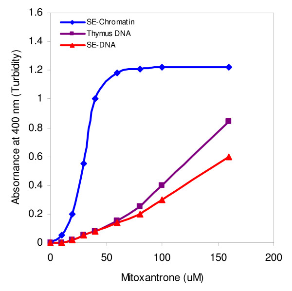
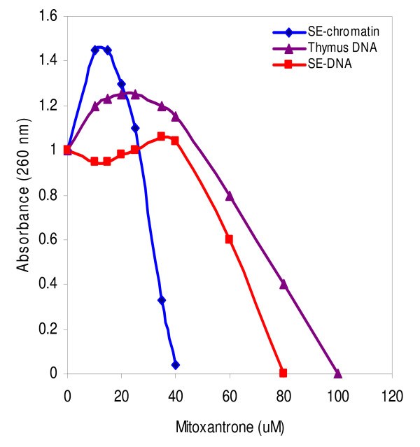
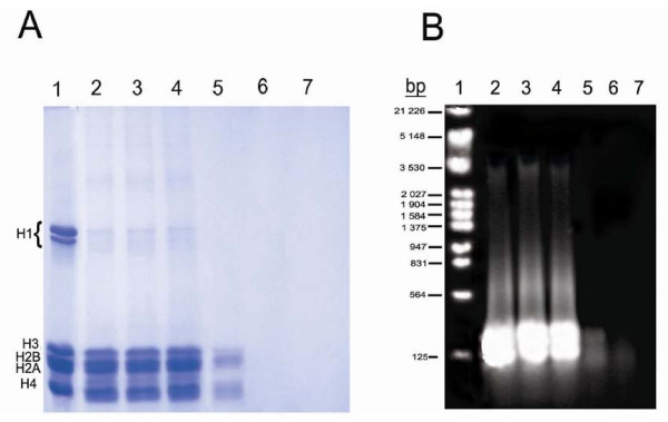
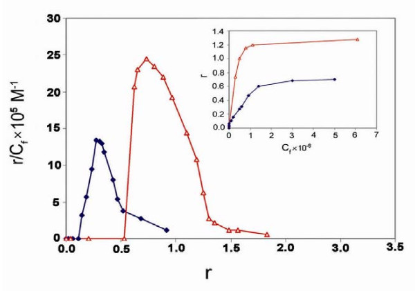
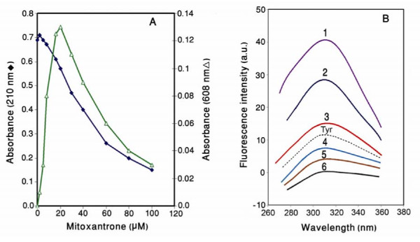
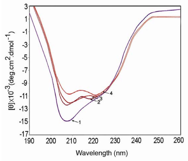
Mitoxantrone is a potent antitumor drug, widely used in the treatment of various cancers. In the present study, we have investigated and compared the affinity of anticancer drug, mitoxantrone, to EDTA-soluble chromatin (SE-chromatin), DNA and histones employing UV/Vis, fluorescence, CD spectroscopy, gel electrophoresis and equilibrium dialysis techniques. The results showed that the interaction of mitoxantrone with SE-chromatin proceeds into compaction/aggregation as revealed by reduction in the absorbencies at 608 and 260 nm (hypochromicity) and disappearance of both histones and DNA on the gels. Mitoxantrone interacts strongly with histone proteins in solution making structural changes in the molecule as shown by CD and fluorescence analysis. The binding isotherms demonstrate a positive cooperative binding pattern for the chromatin- mitoxantrone interaction. It is suggested higher binding affinity of mitoxantrone to chromatin compared to DNA implying that the histone proteins may play an important role in the chromatin- mitoxantrone interaction process.


Figures

Figure 1
Figure 1
Turbidity measurements at 400 nm of the interaction of various concentrations of mitoxantrone with SE-chromatin (diamond), SE-DNA (triangle) and thymus DNA (square). The reaction was carried out in 10 mM Tris-HCl (pH 7.5) and incubation time was 45 min at 23°C. Results are average of 3 individual experiments.
Figure 2
Figure 2
Absorbance changes at 260 nm of the supernatants obtained from the interaction of mitoxantrone with SE-chromatin (diamond), SE-DNA (triangle) and calf thymus DNA (square). Experimental condition was the same as described in figure 1.
Figure 3
Figure 3
A: 15% SDS gel electrophoresis of the supernatants obtained from the interaction of various concentrations of mitoxantrone with SE-chromatin. Lanes 2–7 are 0, 10, 20, 30, 40 and 60 μM of mitoxantrone respectively. Lane 1, calf thymus histones as a standard. B: Agarose gel of DNA extracted from the supernatants. Lane 1, EcoR1 DNA marker. Lanes 2–7 are the same as in gel A. Number of experiments was 3.
Figure 4
Figure 4
Scatchard plots of the binding of mitoxantrone to SE-chromatin (diamond) and DNA (triangle) carried out in 10 mM Tris-HCl pH 7.4 for 72 hours at 25°C. Results are means of 3 individual experiments.
Figure 5
Figure 5
A) Absorbance changes of histones upon mitoxantrone binding. The reaction was carried out in 10 mM Tris-HCl pH 7.3 at 23°C. B) Fluorescence emission spectra of the histones in the presence and absence of various concentrations of mitoxantrone. All samples were prepared in 10 mM Tris – HCl (pH 7.3) and incubation time after drug addition was 20 minutes. Excitation was at 278 nm. Spectra 1–6 are 0, 10, 20, 30, 40, and 60 μM of mitoxantrone respectively. Tyr: tyrosine spectrum given for comparison.
Figure 6
Figure 6
Far UV CD spectra of the histones in 10 mM Tris-HCl at pH 7.3 in the absence and presence of mitoxantrone. Each spectrum was obtained at 25°C with a 10 mm path length cell. 1–4 are 0, 10, 20 and 40 μM of mitoxantrone respectively. All necessary corrections were made for background absorption.

References

    1. Holmes FA, Yap HY, Esparza L, Buzdar AU, Hortobaqui GN, Blumenschein GR. Mitoxantrone, cyclophosphamid and 5-fluorouracil in the treatment of hormonally unresponsive metastatic breast cancer. Semin Oncol. 1984;11:28–31. - PubMed
    1. Velasquez WS, Lew D, Grogan TM, Spiridonidis CH, Balcerzak SP, Dakhil SR, et al. Combination of fludarabine and mitoxantrone in untreated stages III and IV low-grade lymphoma. J Clin Oncol. 2003;21:1996–2003. doi: 10.1200/JCO.2003.09.047. - DOI - PubMed
    1. Thomas X, Archimbaud E. Mitoxantrone in the treatment of acute myelogenous leukemia. Hematol Cell Ther. 1997;39:63–74. doi: 10.1007/s00282-997-0163-8. - DOI - PubMed
    1. White RJ, Durr FF. Development of mitoxantrone. Invest New Drugs. 1985;3:85–93. doi: 10.1007/BF00174154. - DOI - PubMed
    1. Koutinos G, Stathopoulos GP, Dontas I, Perrea-Kotsarolis D, Couris E, Kararannacos PE, et al. The effect of doxorubicin and its analogue mitoxantrone on cardiac muscle and on serum lipids: an experimental study. Anticancer Res. 2002;22:815–20. - PubMed

Publication types