

Cardioprotection by metabolic shut-down and gradual wake-up
- PMID: 19285082
- PMCID: PMC2683198
- DOI: 10.1016/j.yjmcc.2009.02.026
Cardioprotection by metabolic shut-down and gradual wake-up
Abstract
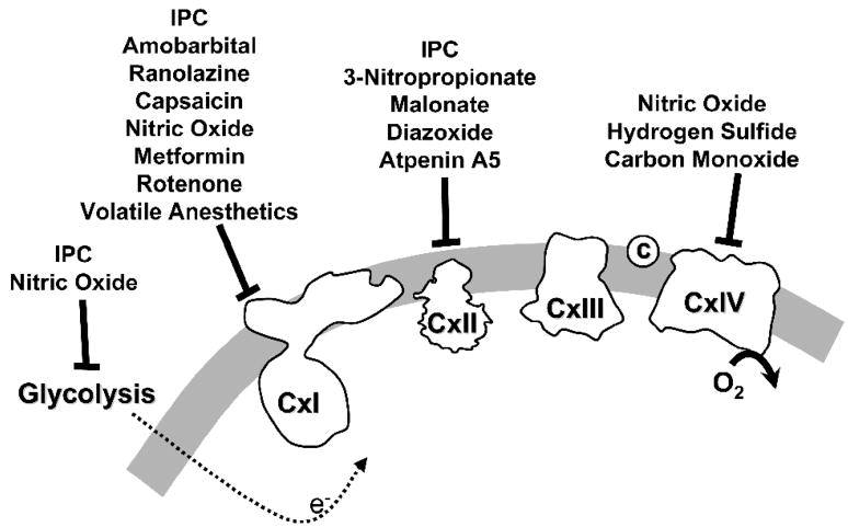
Mitochondria play a critical role in cardiac function, and are also increasingly recognized as end effectors for various cardioprotective signaling pathways. Mitochondria use oxygen as a substrate, so by default their respiration is inhibited during hypoxia/ischemia. However, at reperfusion a surge of oxygen and metabolic substrates into the cell is thought to lead to rapid reestablishment of respiration, a burst of reactive oxygen species (ROS) generation and mitochondrial Ca(2+) overload. Subsequently these events precipitate opening of the mitochondrial permeability transition (PT) pore, which leads to myocardial cell death and dysfunction. Given that mitochondrial respiration is already inhibited during hypoxia/ischemia, it is somewhat surprising that many respiratory inhibitors can improve recovery from ischemia-reperfusion (IR) injury. In addition ischemic preconditioning (IPC), in which short non-lethal cycles of IR can protect against subsequent prolonged IR injury, is known to lead to endogenous inhibition of several respiratory complexes and glycolysis. This has led to a hypothesis that the wash-out of inhibitors or reversal of endogenous inhibition at reperfusion may afford protection by facilitating a more gradual wake-up of mitochondrial function, thereby avoiding a burst of ROS and Ca(2+) overload. This paper will review the evidence in support of this hypothesis, with a focus on inhibition of each of the mitochondrial respiratory complexes.
Figures
References
-
- Brookes PS, Yoon Y, Robotham JL, Anders MW, Sheu SS. Calcium, ATP, and ROS: a mitochondrial love-hate triangle. Am J Physiol Cell Physiol. 2004;287:C817–C833. - PubMed
-
- Di Lisa F, Bernardi P. Mitochondria and ischemia-reperfusion injury of the heart: fixing a hole. Cardiovasc Res. 2006;70:191–9. - PubMed
-
- Karmazyn M. The myocardial sodium-hydrogen exchanger (NHE) and its role in mediating ischemic and reperfusion injury. Keio J Med. 1998;47:65–72. - PubMed
-
- Kim JS, Ohshima S, Pediaditakis P, Lemasters JJ. Nitric oxide: a signaling molecule against mitochondrial permeability transition- and pH-dependent cell death after reperfusion. Free Radic Biol Med. 2004;37:1943–50. - PubMed
Publication types
MeSH terms
Substances
Grants and funding
LinkOut - more resources
Full Text Sources
Other Literature Sources
Miscellaneous
