. 2009 Jul 15;46(4):1209-21.
doi: 10.1016/j.neuroimage.2009.02.044. Epub 2009 Mar 10.

Left posterior parietal cortex participates in both task preparation and episodic retrieval

Affiliations

Left posterior parietal cortex participates in both task preparation and episodic retrieval

Jeffrey S Phillips et al. Neuroimage. .

Abstract

Optimal memory retrieval depends not only on the fidelity of stored information, but also on the attentional state of the subject. Factors such as mental preparedness to engage in stimulus processing can facilitate or hinder memory retrieval. The current study used functional magnetic resonance imaging (fMRI) to distinguish preparatory brain activity before episodic and semantic retrieval tasks from activity associated with retrieval itself. A catch-trial imaging paradigm permitted separation of neural responses to preparatory task cues and memory probes. Episodic and semantic task preparation engaged a common set of brain regions, including the bilateral intraparietal sulcus (IPS), left fusiform gyrus (FG), and the pre-supplementary motor area (pre-SMA). In the subsequent retrieval phase, the left IPS was among a set of frontoparietal regions that responded differently to old and new stimuli. In contrast, the right IPS responded to preparatory cues with little modulation during memory retrieval. The findings support a strong left-lateralization of retrieval success effects in left parietal cortex, and further indicate that left IPS performs operations that are common to both task preparation and memory retrieval. Such operations may be related to attentional control, monitoring of stimulus relevance, or retrieval.


Figures

Figure 1
Figure 1
Top: Overview of trial structure and hypothesized cognitive processes engaged by task preparation and performance. In the preparatory phase, participants were shown either an episodic (“OLD or NEW”) or semantic (“LIVING or NON”) task preparatory cue for 500 ms, followed by a constant fixation interval of 1500 ms. Preparatory activity is thought to reflect cue processing and task-level preparation, including self-reminding of task goals, decision criteria, and response options. Retrieval phase activity is associated with search of and retrieval from declarative memory, as well as decision-making and response processes. Bottom: Neural responses to preparatory cues and memory robes were independently modeled using a catch-trial design. Compound trials (preparatory cue + memory probe) comprise 80% of trials; catch trials (cue only) comprise 20 percent. Inclusion of catch trials allows sufficient variability to separate cue and probe responses in GLM analysis (Ollinger et al., 2001a, b; Shulman et al., 1999; Wheeler et al., 2006).
Figure 2
Figure 2
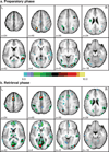
Main effect of time maps from preparatory (top) and retrieval (bottom) phases of experimental trials, indicating regions in which activity significantly changed from baseline. Maps shown are uncorrected for multiple comparisons and thresholded at z>6.0. Regions of interest were defined by masking the uncorrected main effect of time maps with Monte Carlo-corrected, sphericity-adjusted versions of same to exclude peaks which did not meet a corrected alpha-level of 0.05.
Figure 3
Figure 3
Regions of interest defined from preparatory phase main effect of time map. Number labels on timecourse plots indicate the occurrence of statistically significant experimental effects (see legend). Top: significant cue-type differences were observed in pre-SMA, left anterior IPS, left FFG, and right precuneus (Table 6). In the retrieval phase, pre-SMA, left anterior IPS, and left FFG displayed significant task differences. Left anterior IPS additionally exhibited old/new differences. Bottom: bilateral middle and right posterior IPS likewise displayed robust cue responses, although cue-type differences were not statistically significant. All three regions exhibited retrieval phase task differences; additionally, old/new differences were observed in left middle IPS and right posterior IPS. Vertical axis: BOLD signal magnitude as percent change from baseline. Horizontal axis: timecourses extend from 0 to 20 s, beginning from preparatory cue onset; each tick mark corresponds to one 2 s volume of fMRI acquisition. Open stars indicate onset of preparatory cue; closed stars indicate onset of memory probes. Retrieval phase timecourses are based on correct trials only. For visualization purposes, the five parietal ROIs have been projected onto inflated cortical surfaces (Van Essen et al., 2001). Posterior views are displayed in the right panels. Note that contiguous ROIs can become separated in the warping and unfolding procedure. a = anterior, m = middle, p = posterior.
Figure 4
Figure 4
Left parietal regions showing old/new differences, identified from retrieval phase main effect of time map. Number labels on timecourse plots indicate the occurrence of statistically significant experimental effects (see legend). Left inferior precuneus, superior precuneus, and IPL all exhibit significant effects of task and study history in the retrieval phase (Table 6). Vertical axis: BOLD signal magnitude as percent change from baseline. Horizontal axis: timecourses extend from 0 to 20 s, beginning from preparatory cue onset; each tick mark corresponds to one 2 s volume of fMRI acquisition. Open stars indicate onset of preparatory cue; closed stars indicate onset of memory probes. Retrieval phase timecourses are based on correct trials only.
Figure 5
Figure 5
Regions previously implicated in attentional control, identified from retrieval phase main effect of time map. Number labels on timecourse plots indicate the occurrence of statistically significant experimental effects (see legend). All regions except left anterior and posterior DLPFC exhibit significant retrieval phase task differences. Old/new differences occur in left AI and left anterior DLPFC; in all other regions, study history effects are non-significant. Vertical axis: BOLD signal magnitude as percent change from baseline. Horizontal axis: timecourses extend from 0 to 20 s, beginning from preparatory cue onset; each tick mark corresponds to one 2 s volume of fMRI acquisition. Open stars indicate onset of preparatory cue; closed stars indicate onset of memory probes. Retrieval phase timecourses are based on correct trials only.

References

    1. Astafiev SV, Shulman GL, Stanley CM, Snyder AZ, Van Essen DC, Corbetta M. Functional organization of human intraparietal and frontal cortex for attending, looking, and pointing. Journal of Neuroscience. 2003;23:4689–4699. - PMC - PubMed
    1. Balan PF, Gottlieb J. Integration of exogenous input into a dynamic salience map revealed by perturbing attention. Journal of Neuroscience. 2006;26:9239–9249. - PMC - PubMed
    1. Buckner RL. Functional-anatomic correlates of control processes in memory. Journal of Neuroscience. 2003;23:3999–4004. - PMC - PubMed
    1. Buckner RL, Goodman J, Burock M, Rotte M, Koutstaal M, Schacter DL, Rosen B, Dale AM. Functional-anatomic correlates of object priming in humans revealed by rapid presentation event-related fMRI. Neuron. 1998a;20:285–296. - PubMed
    1. Buckner RL, Koutstaal W, Schacter DL, Wagner AD, Rosen BR. Functional-anatomic study of episodic retrieval using fMRI: I Retrieval effort versus retrieval success. Neuroimage. 1998b;7:151–162. - PubMed

Publication types