. 2009 Apr 14;19(7):567-72.
doi: 10.1016/j.cub.2009.02.037. Epub 2009 Mar 12.

Polymorphic members of the lag gene family mediate kin discrimination in Dictyostelium

Affiliations

Polymorphic members of the lag gene family mediate kin discrimination in Dictyostelium

Rocio Benabentos et al. Curr Biol. .

Abstract

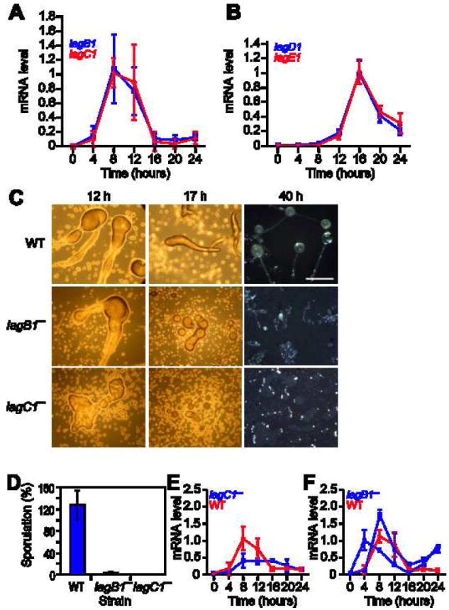
Self and kin discrimination are observed in most kingdoms of life and are mediated by highly polymorphic plasma membrane proteins. Sequence polymorphism, which is essential for effective recognition, is maintained by balancing selection. Dictyostelium discoideum are social amoebas that propagate as unicellular organisms but aggregate upon starvation and form fruiting bodies with viable spores and dead stalk cells. Aggregative development exposes Dictyostelium to the perils of chimerism, including cheating, which raises questions about how the victims survive in nature and how social cooperation persists. Dictyostelids can minimize the cost of chimerism by preferential cooperation with kin, but the mechanisms of kin discrimination are largely unknown. Dictyostelium lag genes encode transmembrane proteins with multiple immunoglobulin (Ig) repeats that participate in cell adhesion and signaling. Here, we describe their role in kin discrimination. We show that lagB1 and lagC1 are highly polymorphic in natural populations and that their sequence dissimilarity correlates well with wild-strain segregation. Deleting lagB1 and lagC1 results in strain segregation in chimeras with wild-type cells, whereas elimination of the nearly invariant homolog lagD1 has no such consequences. These findings reveal an early evolutionary origin of kin discrimination and provide insight into the mechanism of social recognition and immunity.


Figures

Figure 1
Figure 1. Sequence polymorphism
We amplified and sequenced genes from several D. discoideum strains and aligned the sequences of each gene separately. We determined the position of variable nucleotides and determined whether each variation corresponds to a synonymous (S) or a non-synonymous (N) variation in the protein sequence. The upper box of each panel represents the synonymous variations as green bars above the zero-line and the non-synonymous variations as red bars below the line. The x-axis indicates the codon number in the ORF and the y-axis indicates the frequency of the variation in each strain compared to all the other strains. We used the nucleotide variation data to compute the dN/dS ratio at each codon in a sliding window of 31 codons along the entire coding sequence. The data are plotted in the lower box of each panel, the x-axis indicates the codon number in the ORF and the y-axis indicates the dN/dS ratio. Data above the line (dN/dS = 1) suggest that the region is under balancing or positive selection. A. lagB1 exhibited a total of 266 polymorphic codons with as many as 8 variations per codon (30 strains tested). B. lagC1 exhibited 319 polymorphic codons with as many as 7 variants per codon (29 strains tested). We also sequenced lagD1 and lagE1. lagD1 exhibited only 9 polymorphic codons with no more than 2 variants per codon (15 strains tested) and lagE1 exhibited 10 polymorphic codons with only 2 variants per codon (6 strains tested). The dN/dS ratio was much lower than 1 in both cases (data not shown). Information about the strains and the genes we sequenced is provided in Supplement Table S1.
Figure 2
Figure 2. Developmental regulation and function of lag-genes
We used quantitative RT-PCR to measure the mRNA levels of lag-genes in samples collected at 4-hour intervals from developing wild type AX4 cells. At 0 hours, the cells are at the vegetative stage, at 4 hours they are starving, at 8 hours they begin to aggregate, at 12 hours they form tight aggregates with differentiated prespore and prestalk cells, at 16 hours they form fingers with prestalk cells in the anterior end, at 20 hours they begin to culminate and at 24 hours they form mature fruiting bodies with a ball of spores aloft a cellular stalk. The time (hours) is indicated on the x-axis and the mRNA level is indicated on the y-axis in arbitrary units, relative to the maximal level of expression. The results are presented as averages and standard deviations of three technical replications of each of two biological samples (a total of 6 measurements). A. lagB1 mRNA (blue) and lagC1 mRNA (red). B. lagD1 mRNA (blue) and lagE1 mRNA (red). We mutated the lagB1 gene, developed wild type and mutant cells for the indicated time (hours) and analyzed their development. C. Morphological analysis: growth and early developmental properties of the three strains were nearly indistinguishable (data not shown). We show morphological differences between cells developed on non-nutrient agar (12h, 17h) and on dark nitrocellulose filters (40h). The genotypes are indicated on the left. Pictures were taken from above the structure. Bar – 0.5mm. D. Sporulation efficiency: we counted the number of spores collected after 30 hours and present the data as a fraction (%) of the number of amoebae deposited for development. The genotypes are indicated below the bars. Results are the means and standard deviations of 3 independent replications. Gene expression: We used quantitative RT-PCR to measure mRNA levels in samples collected at 4-hour intervals from developing cells. The graphs are as above and the results are presented as averages and standard deviations of three technical replications of each of two biological samples (a total of 6 measurements) except as indicated. E. lagB1 mRNA in wild type cells (red) and in lagC1 cells (blue). F. lagC1 mRNA in wild type cells (red) and in lagB1 cells (blue). The lagB1 cells exhibited large differences between biological samples, probably because the cells do not develop synchronously, so we show 2 biological experiments out of 4 that we have performed (solid and dashed blue lines). The data are averages and standard deviations of three technical replications.
Figure 3
Figure 3. Segregation of cell-cell adhesion mutants from wild type and from lagC1 cells
A. We mixed wild-type (AX4) cells labeled with RFP with different strains labeled with GFP. We developed the cells on a solid substratum, photographed the aggregates after 10-12 hours and after 16-19 hours of development with fluorescence microscopy at the appropriate wavelengths, and merged the red and green images. Bar – 0.5mm. B. To test the adhesion properties of the cells, we developed separately wild-type (AX4) cells labeled with RFP and lagC1 cells labeled with GFP. We disaggregated the cells after 5 hours (B i, bar – 1.0 mm) and after 12 hours (B ii, bar – 0.5 mm) of development, as indicated, mixed the cells with unlabeled counterparts, and allowed them to re-aggregate in shaking suspension. We photographed the mixed aggregates as above. C. To test interactions between pairs of mutant strains, we mixed lagC1 cells labeled with RFP with different strains labeled with GFP (C, i-vi) and lagC1 cells labeled with GFP with different strains labeled with RFP (C, vii-x), as indicated, developed them on a solid substrate and photographed them as above. Bar – 0.5 mm. The genotypes are indicated on the left of each row.

References

    1. Bergelson J, Kreitman M, Stahl EA, Tian D. Evolutionary dynamics of plant R-genes. Science. 2001;292:2281–2285. - PubMed
    1. Boehm T. Quality control in self/nonself discrimination. Cell. 2006;125:845–858. - PubMed
    1. De Tomaso AW, Nyholm SV, Palmeri KJ, Ishizuka KJ, Ludington WB, Mitchel K, Weissman IL. Isolation and characterization of a protochordate histocompatibility locus. Nature. 2005;438:454–459. - PMC - PubMed
    1. Gibbs KA, Urbanowski ML, Greenberg EP. Genetic determinants of self identity and social recognition in bacteria. Science. 2008;321:256–259. - PMC - PubMed
    1. Harada Y, Takagaki Y, Sunagawa M, Saito T, Yamada L, Taniguchi H, Shoguchi E, Sawada H. Mechanism of self-sterility in a hermaphroditic chordate. Science. 2008;320:548–550. - PubMed

Publication types