. 2009 Mar 12;61(5):721-33.
doi: 10.1016/j.neuron.2008.12.025.

Wnt signals organize synaptic prepattern and axon guidance through the zebrafish unplugged/MuSK receptor

Affiliations

Wnt signals organize synaptic prepattern and axon guidance through the zebrafish unplugged/MuSK receptor

Lili Jing et al. Neuron. .

Abstract

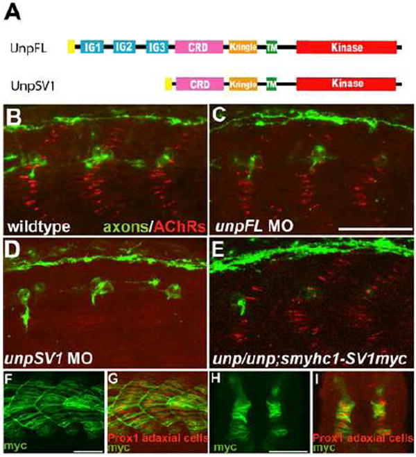
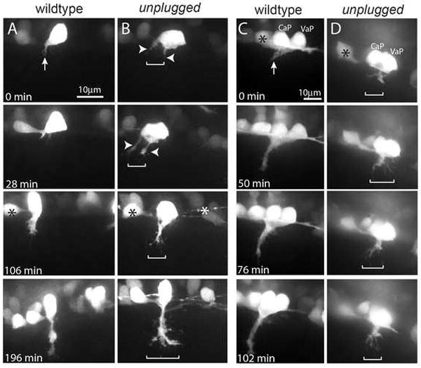
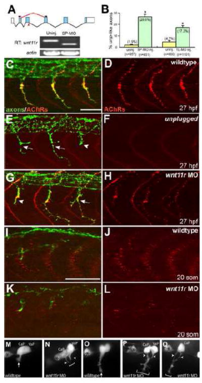
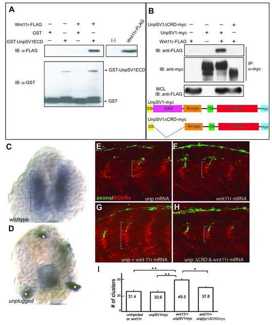
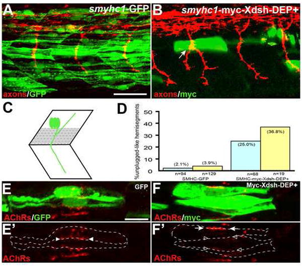
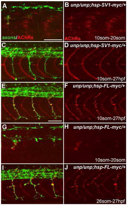
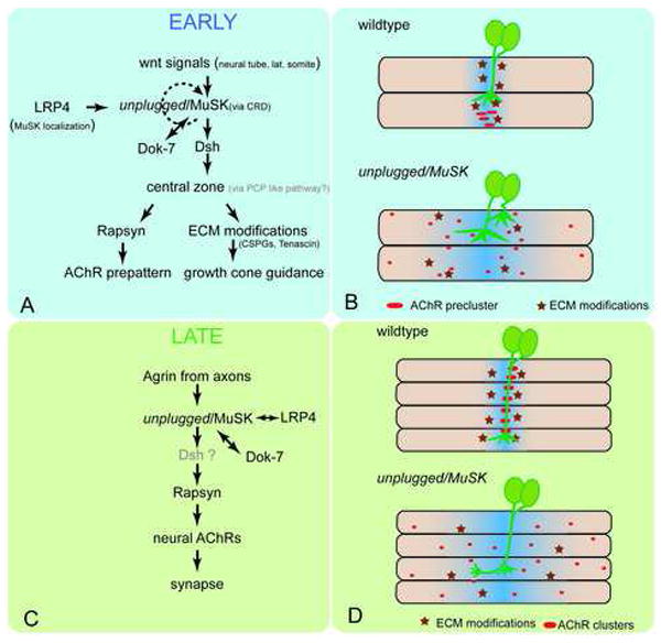
Early during neuromuscular development, acetylcholine receptors (AChRs) accumulate at the center of muscle fibers, precisely where motor growth cones navigate and synapses eventually form. Here, we show that Wnt11r binds to the zebrafish unplugged/MuSK ectodomain to organize this central muscle zone. In the absence of such a zone, prepatterned AChRs fail to aggregate and, as visualized by live-cell imaging, growth cones stray from their central path. Using inducible unplugged/MuSK transgenes, we show that organization of the central muscle zone is dispensable for the formation of neural synapses, but essential for AChR prepattern and motor growth cone guidance. Finally, we show that blocking noncanonical dishevelled signaling in muscle fibers disrupts AChR prepatterning and growth cone guidance. We propose that Wnt ligands activate unplugged/MuSK signaling in muscle fibers to restrict growth cone guidance and AChR prepatterns to the muscle center through a mechanism reminiscent of the planar cell polarity pathway.


Figures

Figure 1
Figure 1. UnpSV1 controls AChR prepatterning
(A) Domain structure of the Unplugged protein isoforms. (B-E) Lateral views of caudal segments in 17 hpf embryos stained for motor axons (green, znp-1/SV2) and AChRs (red, α-BTX). (B) In wildtype embryos, AChRs are prepatterned in a central band along the dorsal and ventral myotome before the first growth cones approach. (C) UnpFL MO injection does not affect AChR prepattern. (D) UnpSV1 MO injection causes complete absence of AChR prepattern. (E) UnpSV1 expression in adaxial cells restores AChR prepattern in unplugged mutant embryos. (F-G) Lateral views and cross-sectional views (H, I) of 17 hpf Tg(smyhc1:UnpSV1myc) embryos stained with anti-myc (green) and anti-Prox1 (red), which labels the nuclei of adaxial cells. Scale bars: 50 μm.
Figure 2
Figure 2. unplugged restricts navigating growth cones to a central muscle zone
(A-D) Still images from time lapse movies showing the initial migration of single CaP axons (A, B), or CaP/VaP pair axons (C, D) from the spinal cord into the myotome. Arrows point to the single wildtype CaP growth cone (A) and to the tightly fasciculated wildtype CaP/VaP growth cones (C). In contrast unplugged CaP neurons form extensive filopodia and even multiple growth cones (arrowheads) that occupy a broader area (brackets, B). Similary, mutant CaP/VaP growth cones appear defasciculated and occupy a broader area compared to wildtype. Asterisks indicate interneurons also labeled by the Tg(Hb9:GFP) transgene.
Figure 3
Figure 3. wnt11r is critical for axonal guidance and AChR prepatterning
(A) The splice morpholino (SP-MO) targets the splice donor site of the wnt11r exon 3 (red arrow), and MO-induced aberrant splicing is shown in red. RT-PCR analyses of uninjected and wnt11r SP-MO injected embryos (arrows indicate the position of PCR primers). (B) Quantification of wnt11r MOs injected embryos. TL-MO, translation initiation mopholino. Per embryo, twenty hemisegments were analyzed; n=hemisegments. Results are expressed as the mean of multiple injection experiments ±s.e.m., (*p<0.001, t test). (C-L) Wildtype, unplugged and wnt11r MO injected embryos at 27hpf (C-H), and at the 20-somite stage (I-L), stained for motor axons (znp-1, green) and AChR clusters (α-BTX, red). (E, F) In contrast to wildtype, unplugged embryos display characteristic stalling (arrowhead) and branches (arrows) at the choice point, and lack all AChR clusters. (G, H) Injection of wnt11r MO causes unplugged like axonal stalling (arrowhead), branching (arrow), and a strong reduction of AChR prepatterning (K, L). Note that the size and intensity of neural AChR clusters is reduced in wnt11r 27 hpf morphants (H). (M-Q) Time-lapse images of Hb9-GFP labeled wildtype (M, O) and wnt11r morphant CaP and VaP axons (N, P, Q), as they exit from the spinal cord (M, N), and as they reach the somitic choice point (O-Q). Asterisks indicate the cell body of interneurons. (M, O) Wildtype CaP and VaP neurons extended one growth cone (arrow). Note the broad area (brackets) the two defasciculated wnt11r morphants CaP/VaP growth cones occupy (arrowheads in N, P, Q), compared to wildtype (M, O). Scale bars: 50 μm.
Figure 4
Figure 4. Wnt11r binds to UnpSV1 and overepressions of wnt11r and unpSV1 increase prepatterning
(A) Binding of Wnt11r to the extracellular domain (ECD) of UnpSV1 in vitro. GST-UnpSV1ECD fusion proteins, coupled to glutathione sepharose, were mixed with conditioned media containing secreted Wnt11r-FLAG. Amounts of GST-UnpSV1 and Wnt11r-FLAG used in analysis were assessed by anti-GST (lower panel) and anti-FLAG (right panel) immunoblotting (IB), respectively. Amounts of Wnt11r-FLAG bound were evaluated by anti-FLAG IB (upper panel). (B) Coimmunoprecipitation of UnpSV1 with Wnt11r in 293T cells. 293T cells were cotransfected with Wnt11r-FLAG and UnpSV1-myc or its CRD deletion mutant. Whole cell lysates (WCL) were subjected to anti-FLAG IB to determine the expression of Wnt11r-FLAG (lower panel). The UnpSV1-bound Wnt11r was assessed by IB of the anti-myc immunoprecipitate (upper panel). Schematic diagrams of constructs used in the experiments. SS: signal sequence. (C and D) Cross sections of 20 somite stage embryos injected with purified Wnt11r-FLAG protein. (C) In wildtype embryos, Wnt11r binds to adaxial cells as highlighted by the brackets. Binding is abolished in unplugged mutants(asterisks in D mark non-specific staining). (E-H) Wildtype embryos were injected with mRNAs as indicated. The domain of AChR prepatterning (brackets) was expanded in embryos co-injected with wnt11r and unpSV1myc mRNAs, and was dependent on the CRD domain (G, H). (I) Co-overexpression of wnt11r and unpSV1 significantly increases the number of prepatterned clusters/hemisegment (n=5-18 hemisegments per bar, average=10). Results are expressed as the average of different injection experiments (t test, **p<0.01, *p<0.05). Amounts of mRNA (ng/embryo): wnt11r-FLAG, 0.3; SV1myc, 0.5; SV1ΔCRDmyc, 0.5. AChR cluster size distribution was not altered.
Figure 5
Figure 5. Inhibition of the non-canonical Dsh pathway in adaxial fibers
(A) Stochastic expression of Tg(smyhc1:GFP) in adaxial muscle (green) does not affect motor axons (red). (B) Expression of Tg(smyhc1:myc-XDsh-DEP+) (green) in adaxial fibers dorsal to the choice point causes unplugged like pathfinding defects (arrow). (C) Location of the dorsal 6-7 adaxial cells (in grey) used for scoring. (D) Analysis of axonal phenotypes. (n=hemisegments; blue, hemisegments with 2 adaxial cells expressing the transgene; yellow, hemisegments with 3 or more adaxail cells expressing the transgene). (E-F’) Confocal images of adjacent adaxial muscle pioneers expressing the smyhc1-GFP or smyhc1-myc-Xdsh-DEP+ transgene. Only AChR clusters between two adjacent transgene positive adaxial cells were analyzed (outlined by dashed lines). Tg(smyhc1:GFP) expressing adaxial cells form AChR clusters (arrowheads in E’), while Tg(smyhc1:myc-XDsh-DEP+ expression disrupts AChR clusters between transgene expressing cells (F’, open arrowhead); note that this does not affect adjacent, non-transgenic cells which formed normal AChR clusters (F’, arrows). For each transgene, four embryos with GFP or Myc-Dsh-DEP+ positive adaxial cells were analyzed. Prepatterned clusters were reduced in all Myc-Dsh-DEP+ expressing embryos. Scale bars: A, 50 μm; E, 10 μm.
Figure 6
Figure 6. Neuromuscular synapses form in the absence of AChR prepattern
20-somite stage (A-B, G-H) or 27 hpf (C-F and I-J) embryos after heat shock treatment. (A, B) Tg(hsp70l:UnpSV1-myc;unplugged) embryos received heat shock from the 10- to 20-somite stage, which rescued AChR prepattern. (C, D) Similar heat shock treatment (10-somite to 27 hpf) also restored motor axon pathfinding, but not neuromuscular synapses. (E, F) The same heat shock treatment rescued motor axons and neuromuscular synapses in Tg(hsp70l:UnpFL-myc; unplugged) embryos. (G, H) In contrast, heat shock between the 10- and 20-somite stage failed to rescue AChR prepattern in Tg(hsp70l:UnpFL-myc;unplugged) embryos. (I, J) Heat shock treatment of same embryos between the 26-somite stage and 27 hpf, i.e. after the time period of prepatterning, was sufficient to rescue neuromuscular synapses. Scale bars: 50 μm.
Figure 7
Figure 7. unplugged/MuSK signaling during synapse formation
Signaling during the early (A, B) and late (C, D) phase of neuromuscular synapse formation. (A, B) Early during synapse formation, Wnt signals act through unplugged/MuSK receptor to establish a central muscle zone, possibly through a Dsh-dependant, PCP like pathway. LRP4 is essential for MuSK localization, and Dok-7 for MuSK activation (Okada et al., 2006). Ligand dependent unplugged/MuSK activation may rapidly become ligand-independent. One branch of this pathway requires rapsyn to cluster AChRs (red ovals in B) in a central prepattern, while through a rapsyn-independent mechanism, e.g. modifications of the ECM components (dark red stars in B), growth cones are restricted to the central zone. In the absence of Wnt or unplugged/MuSK, rapsyn is not activated and thus AChRs are dispersed throughout the muscle and navigating growth cones extend into lateral muscle territory. Blue shades indicate the central zone. (C, D) During the late phase, nerve-derived agrin signals through unplugged/MuSK and LRP4 to recruit rapsyn, which stabilizes neural AChRs and promotes synapse development. (D) In the absence of unplugged/MuSK, rapsyn is not recruited and thus AChR cluster are not stabilized in the central zone. Absence of unplugged also causes rapsyn-independent pathfinding defects, possibly through the lack of ECM modifications. Note that in the absence of a central muscle zone at the early stages, no AChR prepattern forms, but that local agrin secretion from the axon and late expression of unplugged/MuSK appears sufficient to induce neural AChRs and subsequently functional synapses.

References

    1. Anderson MJ, Cohen MW. Nerve-induced and spontaneous redistribution of acetylcholine receptors on cultured muscle cells. J Physiol. 1977;268:757–773. - PMC - PubMed
    1. Axelrod JD, Miller JR, Shulman JM, Moon RT, Perrimon N. Differential recruitment of Dishevelled provides signaling specificity in the planar cell polarity and Wingless signaling pathways. Genes & development. 1998;12:2610–2622. - PMC - PubMed
    1. Bartscherer K, Boutros M. Regulation of Wnt protein secretion and its role in gradient formation. EMBO reports. 2008;9:977–982. - PMC - PubMed
    1. Becker CG, Schweitzer J, Feldner J, Becker T, Schachner M. Tenascin-R as a repellent guidance molecule for developing optic axons in zebrafish. J Neurosci. 2003;23:6232–6237. - PMC - PubMed
    1. Burden SJ. Wnts as retrograde signals for axon and growth cone differentiation. Cell. 2000;100:495–497. - PubMed

Publication types

MeSH terms