

Corneal myofibroblast viability: opposing effects of IL-1 and TGF beta1
- PMID: 19285499
- PMCID: PMC2713809
- DOI: 10.1016/j.exer.2009.03.001
Corneal myofibroblast viability: opposing effects of IL-1 and TGF beta1
Abstract
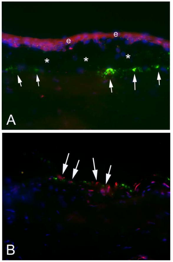
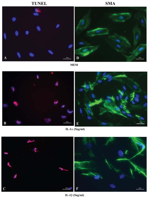
The purpose of this study was to test the effect of corneal epithelial scrape on myofibroblasts associated with haze and elucidate the effect of interleukin-1 and transforming growth factor beta1 on corneal stromal myofibroblasts viability and death in vitro. Corneal epithelial scrape was performed in rabbit eyes with severe haze at one month after -9 diopter photorefractive keratectomy. Corneas were processed for immunocytochemistry for myofibroblast marker alpha-smooth muscle actin (alpha-SMA) and the TUNEL assay to detect apoptosis. Rabbit corneal fibroblasts were cultured with 2 ng/ml of transforming growth factor beta1 (TGF beta1) to induce myofibroblast differentiation confirmed by monitoring alpha-SMA expression. Fluorescence-based TUNEL assay was performed to analyze the apoptotic response of myofibroblasts to IL-1alpha or IL-1beta, in the presence or absence of TGF beta1. Dose response experiments were performed after withdrawal of TGF beta1 and exposure to 1, 5, or 10 ng/ml of IL-1alpha or IL-1beta for 1 h. Subsequent experiments were performed with myofibroblasts exposed to 5 ng/ml of IL-1alpha or IL-1beta in conjunction with 0, 1, 5, or 10 ng/ml of TGF beta1. Corneal epithelial scrape with a scalpel blade produced myofibroblast apoptosis. Exposure to TGF beta1 in vitro resulted in greater than 99% transformation of corneal fibroblasts to alpha-SMA+ myofibroblasts. There was a statistically significant dose-dependent increase in the percentage of TUNEL+ cells with either IL-1alpha or IL-1beta initiated at concentrations as low as 1 ng/ml. For example, after withdrawal of TGF beta1, the % TUNEL+ cells at 1 h after exposure to IL-1alpha increased significantly with increasing concentration (0 ng/ml, 2.4 +/- 0.8% [S.E.M.]; 1 ng/ml, 15.4 +/- 1.8%; 5 ng/ml, 47.4 +/- 3.9%; or 10 ng/ml, 70.3 +/- 3.2%). Similar results were obtained with IL-1beta. The differences between the means of apoptotic myofibroblasts for the different concentrations of cytokine for either IL-1alpha or IL-1beta were significantly different (ANOVA, p < 0.001). When myofibroblasts were exposed to 5 ng/ml of IL-1alpha or IL-1beta, the % TUNEL+ cells at 1 h were reduced in a significant dose-dependent manner when TGF beta1 at a concentration of 5 ng/ml or 10 ng/ml was present in the medium (ANOVA p < 0.01). IL-1alpha or IL-1beta triggers the death of myofibroblasts in vitro and TGF beta1 reduces the IL-1 effect on cell death. TGF beta1 and IL-1 have opposing effects on myofibroblast viability and likely interact to modulate haze generation after corneal injury.
Figures


Similar articles
-
Stromal interleukin-1 expression in the cornea after haze-associated injury.Exp Eye Res. 2010 Sep;91(3):456-61. doi: 10.1016/j.exer.2010.06.023. Epub 2010 Jul 13. Exp Eye Res. 2010. PMID: 20603114 Free PMC article.
-
IL-1 and TGF-β Modulation of Epithelial Basement Membrane Components Perlecan and Nidogen Production by Corneal Stromal Cells.Invest Ophthalmol Vis Sci. 2018 Nov 1;59(13):5589-5598. doi: 10.1167/iovs.18-25202. Invest Ophthalmol Vis Sci. 2018. PMID: 30480706 Free PMC article.
-
Stromal haze, myofibroblasts, and surface irregularity after PRK.Exp Eye Res. 2006 May;82(5):788-97. doi: 10.1016/j.exer.2005.09.021. Epub 2005 Nov 21. Exp Eye Res. 2006. PMID: 16303127 Free PMC article.
-
The corneal fibrosis response to epithelial-stromal injury.Exp Eye Res. 2016 Jan;142:110-8. doi: 10.1016/j.exer.2014.09.012. Exp Eye Res. 2016. PMID: 26675407 Free PMC article. Review.
-
Two-phase mechanism in the treatment of corneal stromal fibrosis with topical losartan.Exp Eye Res. 2024 May;242:109884. doi: 10.1016/j.exer.2024.109884. Epub 2024 Apr 1. Exp Eye Res. 2024. PMID: 38570181 Review.
Cited by
-
Stromal interleukin-1 expression in the cornea after haze-associated injury.Exp Eye Res. 2010 Sep;91(3):456-61. doi: 10.1016/j.exer.2010.06.023. Epub 2010 Jul 13. Exp Eye Res. 2010. PMID: 20603114 Free PMC article.
-
Resolvin E1 analog RX-10045 0.1% reduces corneal stromal haze in rabbits when applied topically after PRK.Mol Vis. 2014 Dec 23;20:1710-6. eCollection 2014. Mol Vis. 2014. PMID: 25558174 Free PMC article.
-
Fibroblastic and bone marrow-derived cellularity in the corneal stroma.Exp Eye Res. 2021 Jan;202:108303. doi: 10.1016/j.exer.2020.108303. Epub 2020 Oct 14. Exp Eye Res. 2021. PMID: 33068626 Free PMC article.
-
Keratocyte Differentiation Is Regulated by NF-κB and TGFβ Signaling Crosstalk.Int J Mol Sci. 2022 Sep 21;23(19):11073. doi: 10.3390/ijms231911073. Int J Mol Sci. 2022. PMID: 36232373 Free PMC article.
-
Hevin plays a pivotal role in corneal wound healing.PLoS One. 2013 Nov 26;8(11):e81544. doi: 10.1371/journal.pone.0081544. eCollection 2013. PLoS One. 2013. PMID: 24303054 Free PMC article.
References
-
- Bonner JC, Lindroos PM, Rice AB, Moomaw CR, Morgan DL. Induction of PDGF receptor-alpha in rat myofibroblasts during pulmonary fibrogenesis in vivo. Am J Physiol Lung Cell Mol Physiol. 1998;274:72–80. - PubMed
-
- Helena MC, Baerveldt F, Kim W-J, Wilson SE. Keratocyte apoptosis after corneal surgery. Invest Ophthalmol Vis Sci. 1998;39:276–83. - PubMed
-
- Jester JV, Huang J, Barry-Lane PA, Kao WW, Petroll WM, Cavanagh HD. Transforming growth factor (beta)- mediated corneal myofibroblast differentiation requires actin and fibronectin assembly. Invest Ophthalmol Vis Sci. 1999b;40:1959–1967. - PubMed
-
- Jester JV, Huang J, Petroll WM, Cavanagh HD. TGF beta induced myofibroblast differentiation of rabbit keratocytes requires synergistic TGF beta, PDGF and integrin signaling. Exp Eye Res. 2002;75:645–657. - PubMed
-
- Jester JV, Petroll WM, Cavanagh HD. Corneal stromal wound healing in refractive surgery: the role of myofibroblasts. Prog Retinal Eye Res. 1999a;18:311–356. - PubMed
Publication types
MeSH terms
Substances
Grants and funding
LinkOut - more resources
Full Text Sources
