. 2009 Jul 18;280(1):31-7.
doi: 10.1016/j.canlet.2009.02.011. Epub 2009 Mar 13.

Naïve human umbilical cord matrix derived stem cells significantly attenuate growth of human breast cancer cells in vitro and in vivo

Affiliations

Naïve human umbilical cord matrix derived stem cells significantly attenuate growth of human breast cancer cells in vitro and in vivo

Rie Ayuzawa et al. Cancer Lett. .

Abstract

The effect of un-engineered (naïve) human umbilical cord matrix stem cells (hUCMSC) on the metastatic growth of MDA 231 xenografts in SCID mouse lung was examined. Three weekly IV injections of 5x10(5) hUCMSC significantly attenuated MDA 231 tumor growth as compared to the saline-injected control. IV injected hUCMSC were detected only within tumors or in close proximity to the tumors. This in vivo result was corroborated by multiple in vitro studies such as colony assay in soft agar and [(3)H]-thymidine uptake. These results suggest that naïve hUCMSC may be a useful tool for cancer cytotherapy.


Figures

Fig. 1
Fig. 1
Effect of hUCMSC on the growth of MDA 231 human breast carcinoma xenografts in CB17 SCID mice. MDA 231 cells (2 × 10⁶ cells) or saline were intravenously inoculated through the tail vein. Eight days after the carcinoma cell inoculation, hUCMSC (5 × 10⁵ cells) were injected through the tail vein. Tumor burden was determined one week after the third weekly hUCMSC treatment. *, p ≤ 0.05 as compared to the untreated MDA 231 alone group.
Fig. 2
Fig. 2
Specific localization of hUCMSC in MDA 231 tumor-bearing mouse lung. One week after three weekly injections of SP-DiI labeled hUCMSC (5 × 10⁵ cells) to MDA 231 tumor-bearing mice, mice were sacrificed and lungs were subjected to immunohistochemical analysis. The fluorescent micrograph shows human mitochondrial stained MDA 231 cells (green), nuclei (blue) stained by Hoechst 33342 and selective engraftment of SP-DiI labeled hUCMS cells (red) in close proximity to the MDA 231 lung tumors (green).
Fig. 3
Fig. 3
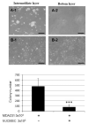
The colony growth of MDA 231 human breast carcinoma cells was significantly attenuated by co-culture with hUCMSC in soft agar. The hUCMSC (3 × 10³) were seeded on the bottom of a 6-well culture dish one day prior to the carcinoma cell seeding. The carcinoma cells (5 × 10⁴) suspended in 0.4% agar were seeded on top of the 0.8% bottom agar layer. Colony growth was evaluated 10 days after the carcinoma cell seeding. The panels A-1and B-1 represent typical views of the MDA 231 colony growth in the middle layer, whereas panels A-2 and B-2 represent views of the control (no cells) and hUCMSC in the bottom of culture dish. Panel C represents a summary of the colony assay. ***, p ≤ 0.0 01 as compared to the colony number of MDA231 alone group.
Fig. 4
Fig. 4
[3H]-Thymidine-uptake into MDA 231 cells was significantly attenuated by co-culture with a small number of hUCMSC. Various numbers of the hUCMSC (0.25, 1.25 and 2.5 × 10⁴) were seeded one day prior to the carcinoma cell (5 × 10⁴) culture in a 12-well culture plate. The [3H] Thymidine-uptake was evaluated 44 hrs after the co-culture, as described in the Methods section. Cell numbers under the horizontal bars indicate hUCMSC co-cultured with MDA 231 cells *, p < 0.05, **, p < 0.01, ***, p < 0.001 as compared to MDA231 alone.
Fig. 5
Fig. 5
Direct co-culture of a small number of hUCMSC and hUCMSC-conditioned medium with MDA 231 cells significantly increased G2 populations in MDA 231 cells as compared with untreated or defined medium treated MDA 231 cells. In the co-culture study, 4 × 10⁴ hUCMSC were seeded one day prior to the 2.4×10⁵ MDA 231 cells. The ratio of the two cell types was 1:6. Three days after co-culture the whole cells were lightly trypsinized and subjected to flow cytometry. Bar graphs indicate the average of two independent triplicate determinations. *, p < 0.05, ***, p ≤ 0.0 01 as compared to the level of untreated control.
Fig. 6
Fig. 6
Direct co-culture of a small number of hUCMSC significantly attenuated Akt (A) and ERK-1/2 phosphorylation (B) in MDA 231 cells, whereas only slightly increased apoptosis-associated signaling of p38 MAPK (C) and PARP cleavage (D) were observed. In the co-culture study, hUCMSC were seeded one day prior to the MDA 231 cells. The ratio of two cell types was 1:30 and 1:6. One day after co-culture the whole cell lysate was prepared and subjected to Western blot analysis. In each panel, the left side picture indicates Western blot analysis, whereas the right side bar graph shows semi-quantification of expressed proteins using scanning densitometry as described in Materials and Methods. The average expression levels normalized by GAPDH level (n=3) are displayed in the histogram.

References

    1. Kuehnle I, Goodell MA. The therapeutic potential of stem cells from adults. BMJ. 2002;325:372–376. - PMC - PubMed
    1. Daar ASS, Sheremeta L. The science of stem cells: Ethical, legal and social issues. Exp. Clin. Transplant. 2003;1:139–146. - PubMed
    1. de Wert G, Mummery C. Human embryonic stem cells: Research, ethics and policy. Hum. Reprod. 2003;18:672–682. - PubMed
    1. Denker H. Embryonic stem cells: An exciting field for basic research and tissue engineering, but also an ethical dilemma. Cells Tissues Organs. 1999;165:246–2495. - PubMed
    1. Henon PR. Human embryonic or adult stem cells: An overview on ethics and perspectives for tissue engineering. Adv. Exp. Med. Biol. 2003;534:27–45. - PubMed

Publication types