. 2009 May 1;388(2):239-51.
doi: 10.1016/j.jmb.2009.03.017. Epub 2009 Mar 13.

Control elements in the neighboring ATPase gene influence spatiotemporal expression of the human agouti-related protein

Affiliations

Control elements in the neighboring ATPase gene influence spatiotemporal expression of the human agouti-related protein

Olha Ilnytska et al. J Mol Biol. .

Abstract

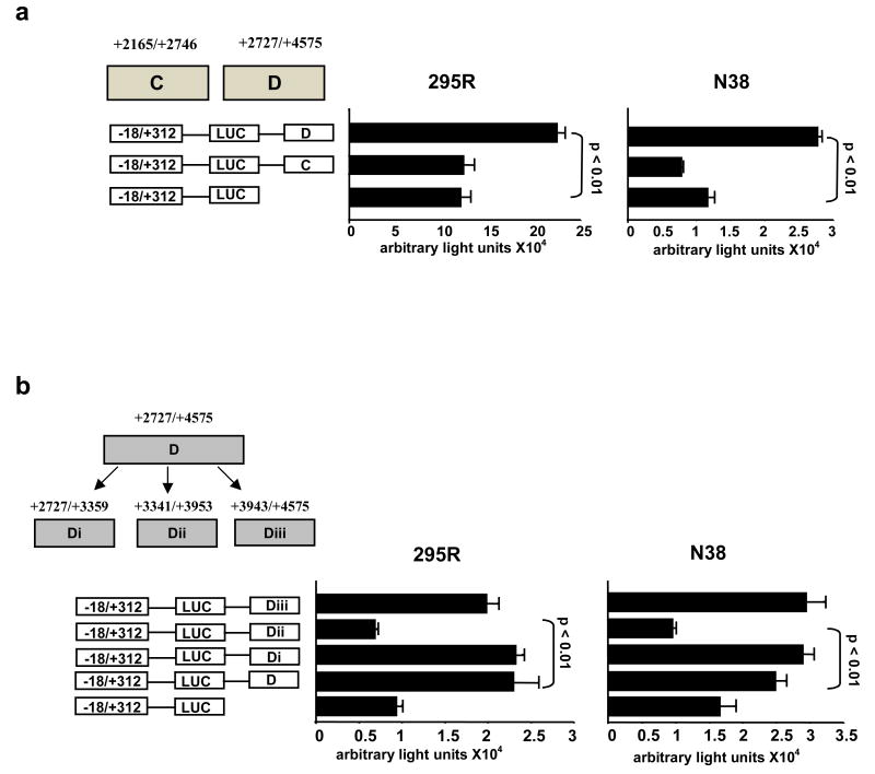
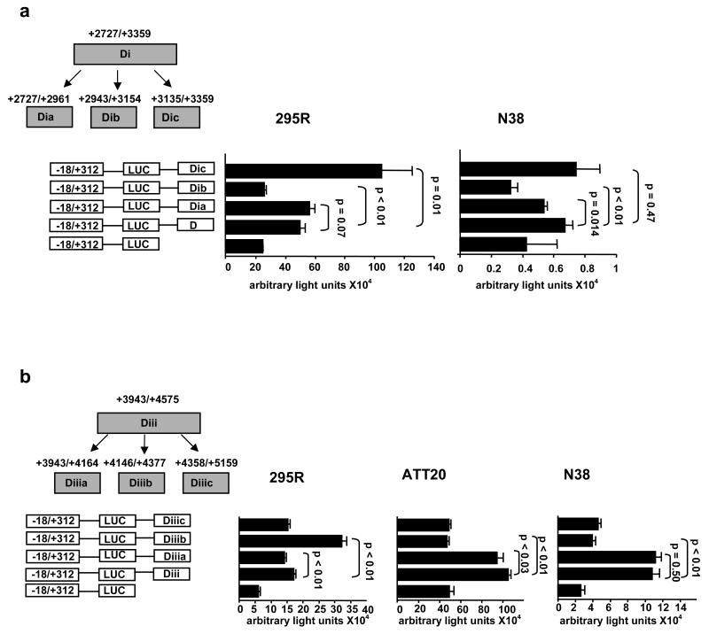
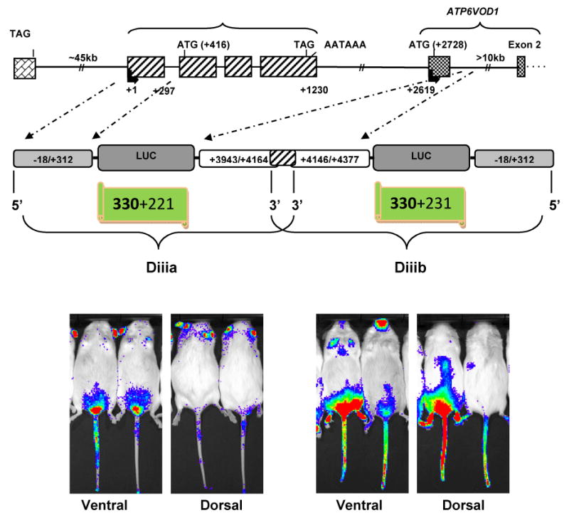
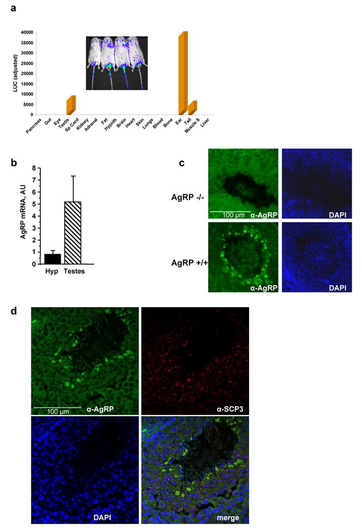
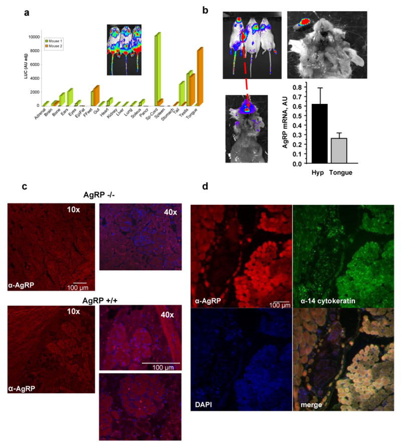
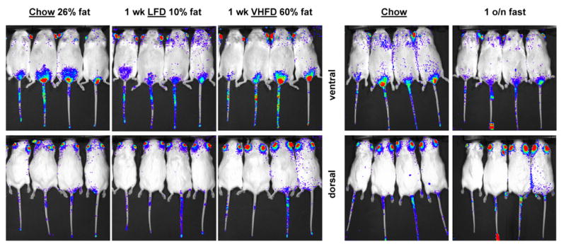
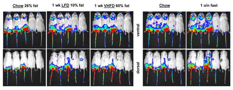
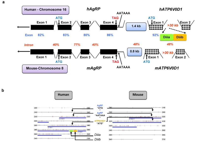
The agouti-related protein (AgRP) is an orexigenic peptide that plays a significant role in the regulation of energy balance. It is expressed in the hypothalamus, the adrenal glands, and the testis, but sequences determining its spatial and temporal expression have not been identified. Using an elaborate in vitro screening approach, we show here that two adjacent enhancers inside the first intron of the neighboring (1.4 kb downstream) ATPase gene (ATP6V0D1) modulate the human AgRP promoter with profound spatiotemporal variation despite their diminutive sizes (221 and 231 nt). In transgenic mice, the proximal enhancer displayed specificity for the testis, tail, and ears, and the distal one for the testis, front feet, bone, heart, muscle, brain, spinal cord, and tongue, while dietary fat and overnight fasting had differential effects on enhancer activities. AgRP in the testis was localized to pachytene spermatocytes and in the tongue to epithelial cells. Comparative sequence analysis showed that the AgRP-ATP6V0D1 intergenic region is two times longer in humans than in mice and that the two enhancers are conserved in the rhesus monkey genome but not in the mouse genome. These data show that spatiotemporal expression of the human AgRP gene is influenced by diversified primate-specific intronic sequences in its neighboring ATP6V0D1 gene.


Figures

Fig. 1
Fig. 1. In vitro determination of enhancer elements
(a) Region D (but not C) increased activity of the human AgRP promoter, equally in the human adrenocortical NCI-h295R and the mouse clonal hypothalamic N38 cell lines. (b) Region D was divided into 3 sub-regions of which only Di and Diii increased basal promoter activity in both cell lines.
Fig. 2
Fig. 2. In vitro elimination of non-enhancer regions
(a) Sub-region Di was divided into smaller portions; Dia, Dib, Dic. Sub-region Dic had the most significant enhancer effect on AgRP basal promoter in both cell lines (b). Sub-region Diii was also divided into smaller regions. Sub-region Diiia had significant enhancer effect in two neuronal cell lines (N38 and ATT20), while sub-region Diiib had significant enhancer effect specifically in the adrenocortical cells (NCI-h295R).
Fig. 3
Fig. 3. Schematic of transgenes and bioluminescence of offspring representing the two enhancers
The upper panel shows a schematic of the AgRP-ATP6V0D1 locus. The middle panel shows schematics of the two constructs (Diiia & Diiib) that were used to make the transgenic mice. The transgene for enhancer Diiib is shown in the reverse orientation (3′ to 5′) so that its exact physical position is represented in the middle panel. The 18 nt overlap of the two enhancers is shown by the shaded region. The bottom panel shows bioluminescence images of dorsal and ventral views from second generation transgenic mice. Images of offspring from the same founder are shown for enhancer Diiia (left, bottom). Images from offspring from two different founders are shown for enhancer Diiib (bottom, right).
Fig. 4
Fig. 4. Luminometric analysis of luciferase signal in Diiia mice and localization of AgRP by immunofluorescence
(a) Luminometric analysis (light units) of luciferase expression in various tissues of Diiia mice (inset shows bioluminescence of a ventral view of these mice). Luciferase is predominantly expressed in the ears, testes, and tails. (b) Real time PCR shows robust expression of AgRP in the testis in comparison to AgRP in the hypothalamus. (c) Immunohistochemistry of mouse testis sections localized AgRP in the middle of seminiferous tubules in wild type (AgRP+/+) but not AgRP-deficient (AgRP-/-) mice. (d) Immunohistochemistry colocalized AgRP with the alpha-synaptonemal complex protein 3 (α-SCP3) in pachytene stage spermatocytes.
Fig. 5
Fig. 5. Luminometric analysis of luciferase signal in Diiib mice and localization of AgRP by immunofluorescence
(a) Luminometric analysis (light units) of luciferase expression in various tissues in Diiib mice (inset shows bioluminescence of a ventral view of these mice). Luciferase is ubiquitously expressed with greater preference for the spinal cord, the tongue, the testis, the ears, the bone, the brain, front feet, soleus muscle, among other tissues. There was significant variation between the two founders indicated by different colors of the bars. (b) Bioluminescence of the signal in the tongue is shown it different zoom modes. Real time PCR shows robust expression of AgRP in the tongue but at lower levels than in the hypothalamus. (c) Immunohistochemistry of mouse tongue sections shows AgRP expression in epithelial cells of wild type (AgRP+/+) but not AgRP-deficient (AgRP-/-) mice. (d) Immunohistochemistry colocalized AgRP with the α-14-cytokeratin that is an epithelial cell-specific protein.
Fig. 6
Fig. 6. In vivo effects of dietary fat and fasting on enhancer Diiia activity
Bioluminescence images of dorsal and ventral views of Diiia offspring while consuming chow, low fat diet (LFD) or very high fat diet (VHFD) for one week each or fasted overnight (o/n) after chow diet. LFD and overnight fasting reduced luciferase expression in a similar fashion.
Fig. 7
Fig. 7. In vivo effects of dietary fat and fasting on enhancer Diiib activity
Bioluminescence images of dorsal and ventral views of Diiib offspring from two founders (first three from Founder A and the other two from Founder B) while consuming chow, low fat diet (LFD) or very high fat diet (VHFD) for one week each or fasted overnight (o/n) after chow diet. LFD and overnight fasting enhanced luciferase expression in a similar fashion. Overnight fasting also led to the appearance of a new signal on the left-hand-side dorsal side in offspring from Founder B.
Fig. 8
Fig. 8. Comparative analysis of human and mouse AgRP-ATP6V0D1 loci
(a) Schematic representation of the human and mouse syntenic AgRP/ATP6V0D1 loci indicating the identity scores (percent) for exons and introns between the two species and presence of enhancers Diiia and Diiib in human only. (b) Algorithmic analysis comparing the CpG islands between the human and mouse syntenic loci encompassing the AgRP-ATP6V0D1 genes. The human AgRP-ATP6V0D1 locus contains three separate CpG islands whereas the mouse locus contains a single continuous CpG island. Enhancers Diiia and Diiib were found to be inside the gap between the second and third CpG islands.

References

    1. Ollmann MM, Wilson BD, Yang YK, Kerns JA, Chen Y, Gantz I, Barsh GS. Antagonism of central melanocortin receptors in vitro and in vivo by agouti-related protein. Science. 1997;278:135–8. - PubMed
    1. Shutter JR, Graham M, Kinsey AC, Scully S, Luthy R, Stark KL. Hypothalamic expression of ART, a novel gene related to agouti, is up- regulated in obese and diabetic mutant mice. Genes Dev. 1997;11:593–602. - PubMed
    1. Ilnytska O, Argyropoulos G. The Role of the Agouti-Related Protein in Energy Balance Regulation. Cell Mol Life Sci 2008 - PMC - PubMed
    1. Pan W, Kastin AJ, Yu Y, Cain CM, Fairburn T, Stutz AM, Morrison C, Argyropoulos G. Selective tissue uptake of agouti-related protein(82-131) and its modulation by fasting. Endocrinology. 2005;146:5533–9. - PubMed
    1. Bewick GA, Gardiner JV, Dhillo WS, Kent AS, White NE, Webster Z, Ghatei MA, Bloom SR. Post-embryonic ablation of AgRP neurons in mice leads to a lean, hypophagic phenotype. Faseb J. 2005;19:1680–2. - PubMed

Publication types

Associated data