. 2009 Mar 19;5(3):234-43.
doi: 10.1016/j.chom.2009.02.005.

Translocation of a Vibrio cholerae type VI secretion effector requires bacterial endocytosis by host cells

Affiliations

Translocation of a Vibrio cholerae type VI secretion effector requires bacterial endocytosis by host cells

Amy T Ma et al. Cell Host Microbe. .

Abstract

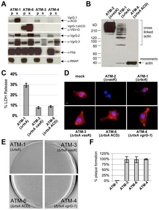
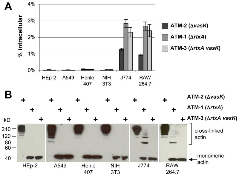
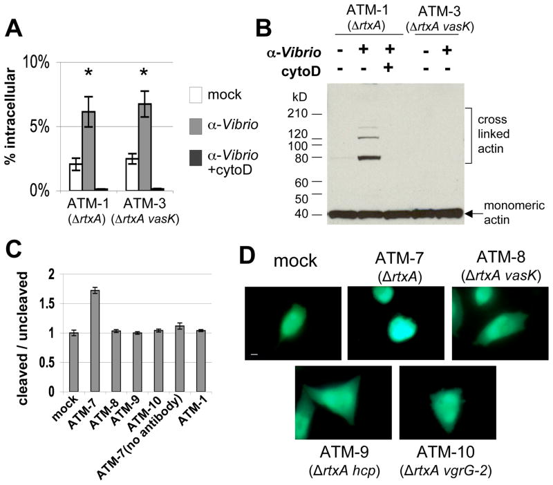
The type VI secretion system (T6SS) is a virulence mechanism common to several Gram-negative pathogens. In Vibrio cholerae, VgrG-1 is required for T6SS-dependent secretion. VgrG-1 is also secreted by T6SS and displays a C-terminal actin crosslinking domain (ACD). Using a heterologous reporter enzyme in place of the ACD, we show that the effector and secretion functions of VgrG-1 are genetically dissociable with the ACD being dispensable for secretion but required for T6SS-dependent phenotypes. Furthermore, internalization of bacteria is required for ACD translocation into phagocytic target cells. Inhibiting bacterial uptake abolishes actin crosslinking, while improving intracellular survival enhances it. Otherwise resistant nonphagocytic cells become susceptible to T6SS-mediated actin crosslinking when engineered to take up bacteria. Our results support a model for translocation of VgrG C-terminal effector domains into target cell cytosol by a process that requires trafficking of bacterial cells into an endocytic compartment where translocation is triggered by an unknown signal.


Figures

Figure 1
Figure 1. The ACD of VgrG-1 is required for host cell phenotypes
A. Western blots of pellet and supernatant fractions of various V52 strains. B. Western blot of host cell actin from J774 cells incubated with various V52 strains at an MOI of 10 for 2 hours. C. Supernatants of J774 cells were analyzed for LDH release one day after exposure to V. cholerae. Values are from triplicate wells and are expressed as percentage of LDH released from lysed mock-treated cells. Error bars indicate +/− one standard deviation. D. Fluorescence microscopy of cells visualizing actin with rhodamine-phalloidin (red) and nuclei with DAPI (blue). Scale bar indicates 10 μm. E. Dictyostelium discoideum plaque assay with various V52 strains. F. Quantification of plaque formation normalized to plaque formation on K. aerogenes.
Figure 2
Figure 2. T6SS mediated actin cross-linking correlates with endocytic uptake into host cells
A. Gentamicin protection assay of various host cell lines exposed to various V52 strains at an MOI of 10 for 1 hour, followed by gentamicin treatment and enumeration of intracellular bacteria. Values are of triplicate wells and are expressed as percentage of initial inoculum recovered. Error bars indicate +/− 1 standard deviation. B. Actin cross-linking in host cells detected by western blot after exposure to various V52 strains at an MOI of 10 for 2 hours.
Figure 3
Figure 3. Cytochalasin D inhibits uptake into J774 cells and in vivo actin crosslinking
A. Western blot against actin from J774 cells in the presence or absence of cytochalasin D. Exposure to V. cholerae was at an MOI of 10 for 2 hours. B. Gentamicin protection assays were performed to verify inhibition of uptake. One hour exposure to V. cholerae at an MOI of 10 was followed by gentamicin treatment and enumeration of intracellular bacteria. Values are of triplicate wells and are expressed as percentage of initial inoculum recovered. Error bars indicate +/− 1 standard deviation. C. In vitro actin cross-linking assay with purified VgrG-1 and actin with various concentrations of cytochalasin D or 0.1% DMSO. After incubation at 37°C for 1 hour, reactions were ran out on a 4–15% gradient SDS-PAGE gel and monitored by western blot.
Figure 4
Figure 4. Bafilomycin A increases ATM-1 induced actin cross-linking in J774 cells
A. Time course of J774 cells exposed to ATM-1 at an MOI of 1 in the presence or absence of bafilomycin A. Actin was visualized by western blot. B. Gentamicin protection assay of J774 in the presence or absence of bafilomycin A and with the addition of cytochalasin D. One hour long incubations at an MOI of 10 were followed by gentamicin treatment and enumeration of intracellular bacteria. Values are of triplicate wells and are expressed as percentage of initial inoculum recovered. Error bars indicate +/− 1 standard deviation. Values between different treatment conditions are statistically significant by Student’s t-test (p < 0.05). C. Actin cross-linking in host cells in the presence of 0.1% DMSO, bafilomycin A alone or bafilomycin A with cytochalasin D. Exposures at MOI of 10 and were 2 hours long. Actin was visualized by western blot. D. In vitro actin cross-linking assay with purified VgrG-1 and actin with various concentrations of bafilomycin A or 0.1% DMSO. After incubation at 37°C for 1 hour, reactions were ran out on a 4–15% gradient SDS-PAGE gel and monitored by western blot.
Figure 5
Figure 5. Translocation of VgrG-1-Bla or VgrG-1ΔACD-Bla into J774 cells
A. Western blot against β-lactamase of pellet and supernatant fractions of various V52 strains producing VgrG-1-Bla or VgrG-1ΔACD-Bla. B. In vitro CCF2-FA cleavage by supernatants of various V52 strains grown in triplicate. Fluorescence was measured with an excitation wavelength of 405 nm and emission wavelengths of 460 nm (cleaved blue channel) and 530 nm (uncleaved green channel). Values are expressed as ratios of cleaved signal to uncleaved signal. Error bars indicate +/− one standard deviation. C. Translocation of VgrG-1-Bla or VgrG-1ΔACD-Bla into J774 cells incubated with various V52 strains. Cells were pre-treated with bafilomycin A, incubated with V52 strains for 2 hours at an MOI of 50, and then loaded with CCF2/AM. Cells were harvested and analyzed on a fluorescence plate reader as for in vitro CCF2-FA assay. Ratios were normalized to mock and error bars indicate +/− 1 standard deviation. D. Gentamicin protection assay on host cells incubated with V. cholerae as for translocation assay and subsequent treated with gentamicin and enumeration of intracellular bacteria. Values are of triplicate wells and are expressed as percentage of initial inoculum recovered. Error bars indicate +/− 1 standard deviation. E. Fluorescence microscopy of J774 cells loaded with CCF2/AM. Scale bar indicates 10 μm. ATM-7, ATM-8, ATM-9, ATM-10 all contain the vgrG-1-bla fusion gene and ATM-11 contains the vgrG-1-ΔACD-bla gene.
Figure 6
Figure 6. Opsonophagocytosis by CHO-FcRγII cells induces translocation of VgrG- 1
A. Gentamicin protection assay of CHO-FcγRII cells incubated with various V52 strains in the presence or absence of α-Vibrio antibody and with cytochalasin D. Values are expressed as percentage of initial inoculum recovered and error bars indicate +/− 1 standard deviation. Groups marked with asterisks are significantly higher than remaining groups (Student’s t-test, p<0.05). B. Western blot against actin from CHO-FcγRII cells incubated with various V52 strains at an MOI of 50 for 30 minutes under various conditions, then treated with gentamicin for an additional 1.5 hours. Actin was visualized by western blot. C. Translocation of VgrG-1-Bla into CHO-FcγRII cells by various V52 strains. Cells were pre-treated with α-Vibrio antibody, incubated with V. cholerae at an MOI of 50 for 2 hours, loaded with CCF2/AM, then harvested for quantification. Fluorescence was measured with an excitation wavelength of 405 nm and emission wavelengths of 460 nm (cleaved blue channel) and 530 nm (uncleaved green channel). Values are expressed as ratios of cleaved signal to uncleaved signal and are normalized to mock. Error bars indicate +/− one standard deviation. D. Fluorescence microscopy of CHO-FcγRII cells incubated with various V52 strains and then loaded with CCF2/AM. Scale bar indicates 10 μm. ATM-7, ATM-8, ATM-9, ATM-10 all encode vgrG-1-bla fusion gene.

Comment in

References

    1. Abd H, Weintraub A, Sandstrom G. Intracellular survival and replication of Vibrio cholerae O139 in aquatic free-living amoebae. Environ Microbiol. 2005;7:1003–1008. - PubMed
    1. Aschtgen MS, Bernard CS, De Bentzmann S, Lloubes R, Cascales E. SciN is an outer membrane lipoprotein required for type VI secretion in enteroaggregative Escherichia coli. J Bacteriol. 2008;190:7523–7531. - PMC - PubMed
    1. Aubert DF, Flannagan RS, Valvano MA. A novel sensor kinase-response regulator hybrid controls biofilm formation and type VI secretion system activity in Burkholderia cenocepacia. Infect Immun. 2008;76:1979–1991. - PMC - PubMed
    1. Barbieri JT, Riese MJ, Aktories K. Bacterial toxins that modify the actin cytoskeleton. Annu Rev Cell Dev Biol. 2002;18:315–344. - PubMed
    1. Bingle LE, Bailey CM, Pallen MJ. Type VI secretion: a beginner’s guide. Curr Opin Microbiol. 2008;11:3–8. - PubMed

Publication types