

Dendritic cells as controllers of antigen-specific Foxp3+ regulatory T cells
- PMID: 19286352
- PMCID: PMC2680740
- DOI: 10.1016/j.jdermsci.2009.02.001
Dendritic cells as controllers of antigen-specific Foxp3+ regulatory T cells
Abstract
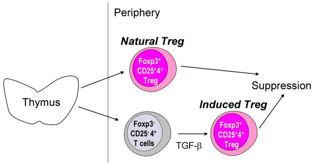
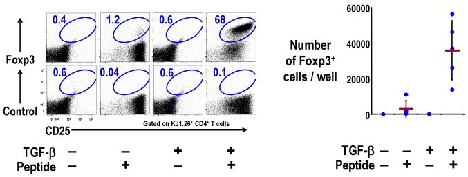
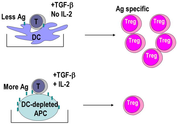
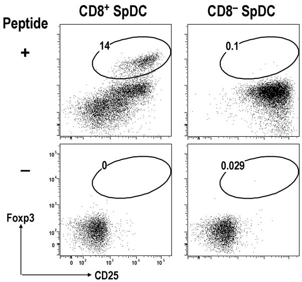
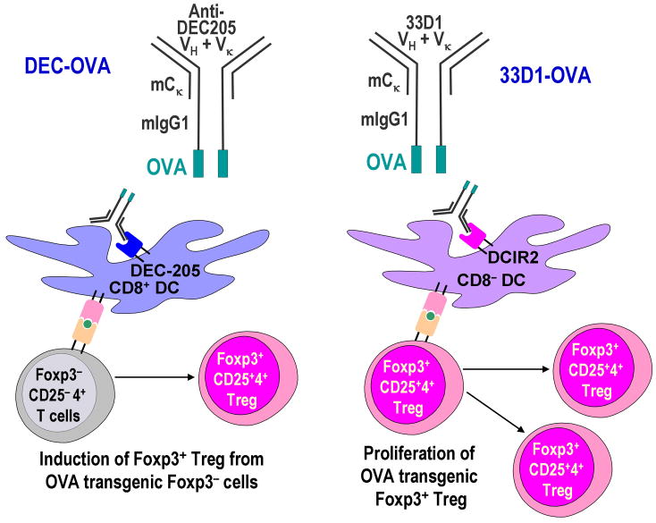
Regulatory T cells (Treg) are a subpopulation of CD4(+) lymphocytes that maintain immunological self-tolerance in the periphery. Treg also regulate or suppress other classes of immune response such as allograft rejection, allergy, tumor immunity, and responses to microbes. Treg express the Foxp3 transcription factor and CD25, the high affinity interleukin-2 receptor (IL-2R). Treg are divided into two types: naturally occurring Treg derived from thymus (natural Treg) and Treg induced from Foxp3(-) CD4(+) T cells in the periphery (induced Treg). It would be valuable to understand how to control the generation of antigen-specific Treg, which could also provide a new approach to treat autoimmunity, allergy or allograft rejection without suppressing immune responses to tumor and microbes. In this review, we will discuss the role of dendritic cells (DCs) in controlling antigen-specific natural Treg and induced Treg. Natural Treg are anergic upon T cell receptor stimulation generally, however, we found that the antigen-specific natural Treg can be expanded by antigen-presenting mature bone marrow-derived dendritic cells (BM-DCs). Furthermore, recent studies showed that antigen-specific Treg can be induced from Foxp3(-) CD25(-) CD4(+) T cells by antigen-presenting DCs, particularly select subsets of DCs in the periphery. These findings need to be pursued to develop novel immune suppressive therapies using antigen-specific Treg educated by DCs.
Conflict of interest statement
Conflict of Interest Statement
Sayuri Yamazaki has no conflicting financial interests. Ralph M Steinman has financial interests in Celldex, which is developing anti–DEC-205 antibodies for human use.
Figures
References
-
- Sakaguchi S. Naturally arising CD4+ regulatory T cells for immunologic self-tolerance and negative control of immune responses. Annu Rev Immunol. 2004;22:531–62. - PubMed
-
- Sakaguchi S, Yamaguchi T, Nomura T, Ono M. Regulatory T cells and immune tolerance. Cell. 2008;133:775–87. - PubMed
-
- Zheng Y, Rudensky AY. Foxp3 in control of the regulatory T cell lineage. Nat Immunol. 2007;8:457–62. - PubMed
-
- Wildin RS, Ramsdell F, Peake J, et al. X-linked neonatal diabetes mellitus, enteropathy and endocrinopathy syndrome is the human equivalent of mouse scurfy. Nat Genet. 2001;27:18–20. - PubMed
-
- Bennett CL, Christie J, Ramsdell F, et al. The immune dysregulation, polyendocrinopathy, enteropathy, X-linked syndrome (IPEX) is caused by mutations of FOXP3. Nat Genet. 2001;27:20–1. - PubMed
Publication types
MeSH terms
Substances
Grants and funding
LinkOut - more resources
Full Text Sources
Other Literature Sources
Research Materials
