

Toward an atomic model of the 26S proteasome
- PMID: 19286367
- PMCID: PMC2743420
- DOI: 10.1016/j.sbi.2009.02.004
Toward an atomic model of the 26S proteasome
Abstract
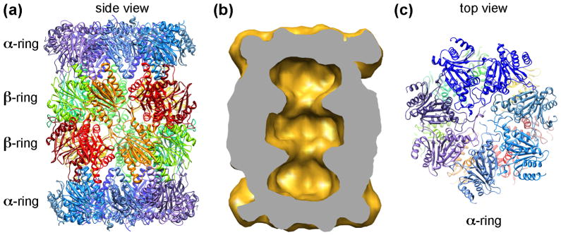
Since the discovery of the 26S proteasome, much progress has been made in determining the structure of this large dynamic protein complex. Until now, a vast amount of structural information of the proteasome has been obtained from all kinds of structure determination techniques, and the function of the protease core is well understood at atomic detail. Yet our understanding of the entire 26S proteasome structure, particularly its 19S regulatory complex, is still limited at a low-resolution blob-ology level. In this review, we highlight the recent progress made in understanding the mechanism of 20S gate opening by the proteasomal activators. We also emphasized the recent methodological advances, particularly in achieving the near atomic resolution by single particle electron cryomicroscopy, and the possible approaches that will enable more detailed structural analysis of the entire 26S proteasome.
Figures
References
-
- Goldberg AL. Nobel committee tags ubiquitin for distinction. Neuron. 2005;45:339–344. - PubMed
-
- Hershko A, Ciechanover A. The ubiquitin system. Annu Rev Biochem. 1998;67:425–479. - PubMed
-
- Ikai A, Nishigai M, Tanaka K, Ichihara A. Electron microscopy of 26 S complex containing 20 S proteasome. FEBS Lett. 1991;292:21–24. - PubMed
-
- Groll M, Ditzel L, Lowe J, Stock D, Bochtler M, Bartunik HD, Huber R. Structure of 20S proteasome from yeast at 2.4 A resolution. Nature. 1997;386:463–471. - PubMed
-
- Lowe J, Stock D, Jap B, Zwickl P, Baumeister W, Huber R. Crystal structure of the 20S proteasome from the archaeon T. acidophilum at 3.4 A resolution. Science. 1995;268:533–539. - PubMed
Publication types
MeSH terms
Substances
Grants and funding
LinkOut - more resources
Full Text Sources
