

Tracking early autoimmune disease by bioluminescent imaging of NF-kappaB activation reveals pathology in multiple organ systems
- PMID: 19286564
- PMCID: PMC2671367
- DOI: 10.2353/ajpath.2009.080700
Tracking early autoimmune disease by bioluminescent imaging of NF-kappaB activation reveals pathology in multiple organ systems
Abstract
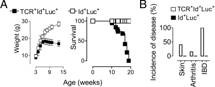
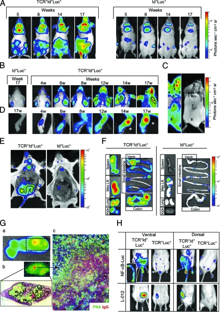
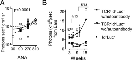
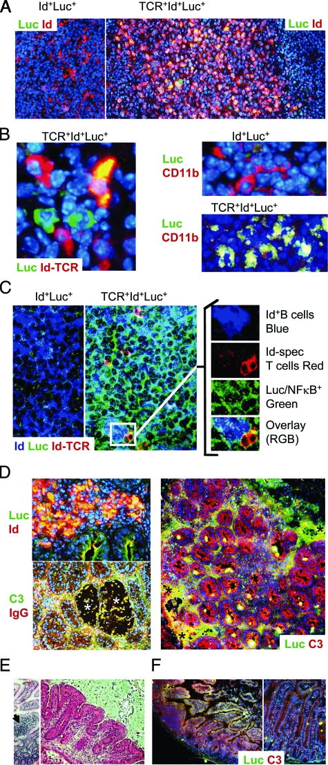
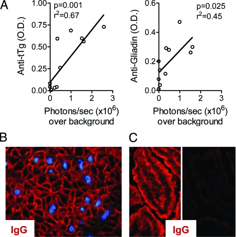
It is desirable to have an early and sensitive detection marker of autoimmune disease in intact animals. Nuclear factor (NF)-kappaB is a transcription factor that is associated with inflammatory responses and immune disorders. Previously, we demonstrated that so-called idiotypic-driven T-B cell collaboration in mice doubly transgenic for paired immunoglobulin and T cell receptor transgenes resulted in a systemic autoimmune disease with systemic lupus erythematosus-like features. Here, we investigated NF-kappaB activation by including an NF-kappaB-responsive luciferase reporter transgene in this animal model. Triply transgenic mice developed bioluminescence signals from diseased organs before onset of clinical symptoms and autoantibody production, and light emissions correlated with disease progression. Signals were obtained from secondary lymphoid organs, inflamed intestines, skin lesions, and arthritic joints. Moreover, bioluminescence imaging and immunohistochemistry demonstrated that a minority of mice suffered from an autoimmune disease of the small intestine, in which light emissions correlated with antibodies against tissue transglutaminase and gliadin. Detection of luciferase by immunohistochemistry revealed NF-kappaB activation in collaborating B and T cells, as well as in macrophages. These results demonstrate that bioluminescent in vivo imaging of NF-kappaB activation can be used for early and sensitive detection of autoimmune disease in an experimental mouse model, offering new possibilities for the evaluation of anti-inflammatory drugs.
Figures

Similar articles
-
Noninvasive nuclear factor-kappaB bioluminescence imaging for the assessment of host-biomaterial interaction in transgenic mice.Biomaterials. 2007 Oct;28(30):4370-7. doi: 10.1016/j.biomaterials.2007.07.005. Epub 2007 Jul 23. Biomaterials. 2007. PMID: 17645941
-
Dynamic imaging of pancreatic nuclear factor κB (NF-κB) activation in live mice using adeno-associated virus (AAV) infusion and bioluminescence.J Biol Chem. 2015 May 1;290(18):11309-20. doi: 10.1074/jbc.M115.647933. Epub 2015 Mar 23. J Biol Chem. 2015. PMID: 25802340 Free PMC article.
-
Cellular immune reaction in the pancreas is induced by constitutively active IkappaB kinase-2.Gut. 2007 Feb;56(2):227-36. doi: 10.1136/gut.2005.084665. Epub 2006 Jul 26. Gut. 2007. PMID: 16870717 Free PMC article.
-
Molecular imaging of the transcription factor NF-kappaB, a primary regulator of stress response.Mutat Res. 2004 Jul 13;551(1-2):199-211. doi: 10.1016/j.mrfmmm.2004.02.024. Mutat Res. 2004. PMID: 15225593 Review.
-
T and B cell tolerance and responses to viral antigens in transgenic mice: implications for the pathogenesis of autoimmune versus immunopathological disease.Immunol Rev. 1991 Aug;122:133-71. doi: 10.1111/j.1600-065x.1991.tb00601.x. Immunol Rev. 1991. PMID: 1937540 Review.
Cited by
-
Making the brain glow: in vivo bioluminescence imaging to study neurodegeneration.Mol Neurobiol. 2013 Jun;47(3):868-82. doi: 10.1007/s12035-012-8379-1. Epub 2012 Nov 29. Mol Neurobiol. 2013. PMID: 23192390 Review.
-
Transgenic Animal Models to Visualize Cancer-Related Cellular Processes by Bioluminescence Imaging.Front Pharmacol. 2019 Mar 15;10:235. doi: 10.3389/fphar.2019.00235. eCollection 2019. Front Pharmacol. 2019. PMID: 30930779 Free PMC article. Review.
-
B cell receptor ligation induces display of V-region peptides on MHC class II molecules to T cells.Proc Natl Acad Sci U S A. 2019 Dec 17;116(51):25850-25859. doi: 10.1073/pnas.1902836116. Epub 2019 Dec 3. Proc Natl Acad Sci U S A. 2019. PMID: 31796587 Free PMC article.
-
iNOS- and NOX1-dependent ROS production maintains bacterial homeostasis in the ileum of mice.Mucosal Immunol. 2018 May;11(3):774-784. doi: 10.1038/mi.2017.106. Epub 2017 Dec 6. Mucosal Immunol. 2018. PMID: 29210363
-
Microbial colonization induces dynamic temporal and spatial patterns of NF-κB activation in the zebrafish digestive tract.Gastroenterology. 2011 Jul;141(1):197-207. doi: 10.1053/j.gastro.2011.03.042. Epub 2011 Mar 24. Gastroenterology. 2011. PMID: 21439961 Free PMC article.
References
-
- Williams WM, Staines NA, Muller S, Isenberg DA. Human T cell responses to autoantibody variable region peptides. Lupus. 1995;4:464–471. - PubMed
-
- Dayan M, Segal R, Sthoeger Z, Waisman A, Brosh N, Elkayam O, Eilat E, Fridkin M, Mozes E. Immune response of SLE patients to peptides based on the complementarity determining regions of a pathogenic anti-DNA monoclonal antibody. J Clin Immunol. 2000;20:187–194. - PubMed
-
- van Schooten WC, Devereux D, Ho CH, Quan J, Aguilar BA, Rust CJ. Joint-derived T cells in rheumatoid arthritis react with self-immunoglobulin heavy chains or immunoglobulin-binding proteins that copurify with immunoglobulin. Eur J Immunol. 1994;24:93–98. - PubMed
-
- Holmoy T, Fredriksen AB, Thompson KM, Hestvik AL, Bogen B, Vartdal F. Cerebrospinal fluid T cell clones from patients with multiple sclerosis: recognition of idiotopes on monoclonal IgG secreted by autologous cerebrospinal fluid B cells. Eur J Immunol. 2005;35:1786–1794. - PubMed
Publication types
MeSH terms
Substances
LinkOut - more resources
Full Text Sources
Other Literature Sources
Medical
Molecular Biology Databases
