. 2009 May;191(10):3237-47.
doi: 10.1128/JB.01837-08. Epub 2009 Mar 13.

Mechanism for sortase localization and the role of sortase localization in efficient pilus assembly in Enterococcus faecalis

Affiliations

Mechanism for sortase localization and the role of sortase localization in efficient pilus assembly in Enterococcus faecalis

Kimberly A Kline et al. J Bacteriol. 2009 May.

Abstract

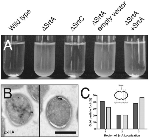
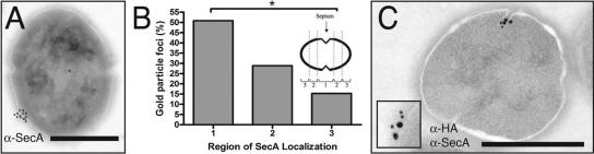
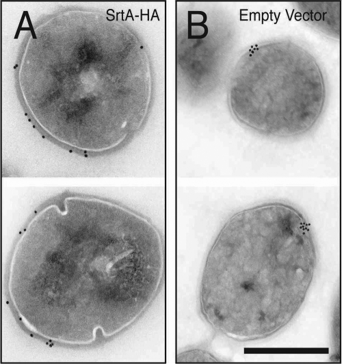
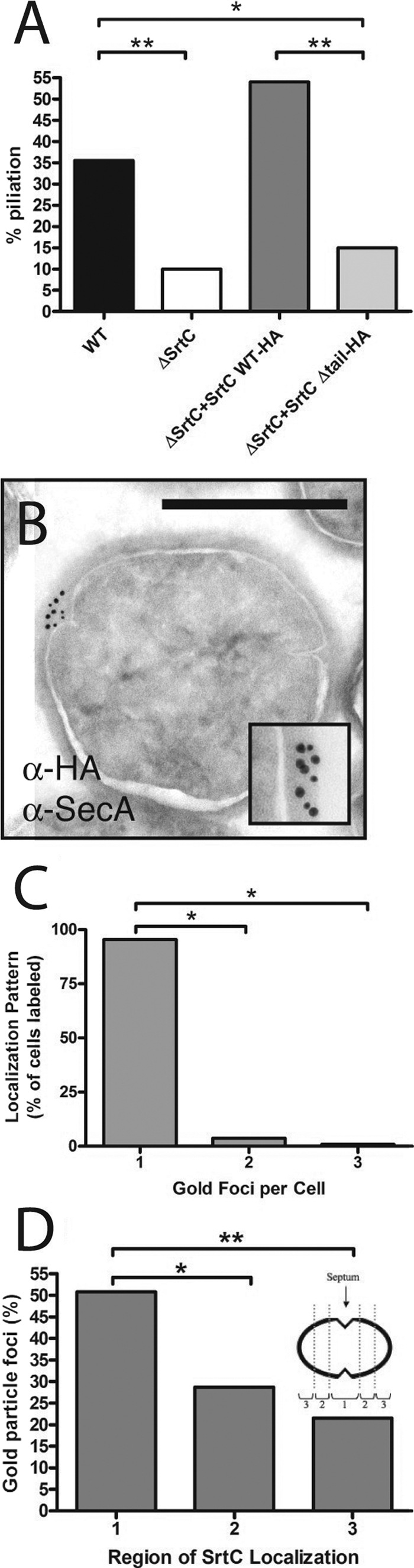
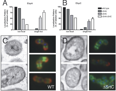
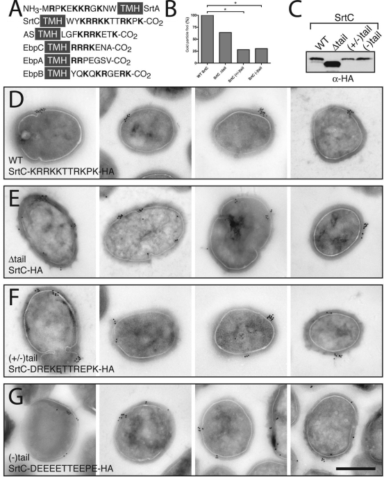
Pathogenic streptococci and enterococci primarily rely on the conserved secretory (Sec) pathway for the translocation and secretion of virulence factors out of the cell. Since many secreted virulence factors in gram-positive organisms are subsequently attached to the bacterial cell surface via sortase enzymes, we sought to investigate the spatial relationship between secretion and cell wall attachment in Enterococcus faecalis. We discovered that sortase A (SrtA) and sortase C (SrtC) are colocalized with SecA at single foci in the enterococcus. The SrtA-processed substrate aggregation substance accumulated in single foci when SrtA was deleted, implying a single site of secretion for these proteins. Furthermore, in the absence of the pilus-polymerizing SrtC, pilin subunits also accumulate in single foci. Proteins that localized to single foci in E. faecalis were found to share a positively charged domain flanking a transmembrane helix. Mutation or deletion of this domain in SrtC abolished both its retention at single foci and its function in efficient pilus assembly. We conclude that this positively charged domain can act as a localization retention signal for the focal compartmentalization of membrane proteins.


Figures

FIG. 1.
FIG. 1.
Sortase A is focally localized in E. faecalis. (A) Broth clumping assay on OG1SS/pCF10-derived strains. Proper sorting of AS and subsequent clumping of E. faecalis is dependent on SrtA but not SrtC. In strains containing SrtA (first, third, and fifth tubes) the bacteria form large aggregates that settle to the bottom of the tube. Strains that lack SrtA (second and fourth tubes) remain turbid. (B) Anti-HA immunoelectron microscopy on OG1SS/pCF10 ΔSrtA complemented with an HA epitope-labeled SrtA expressed under its native promoter. SrtA-HA localizes to single domains on the surface of the bacterium. (C) Quantitative localization analysis of SrtA immunoelectron micrographs. Bacteria were equally divided into three regions from youngest visible septum to pole. Bacteria without a visible septum were excluded from this analysis. Dark gray bars represent log-phase bacteria, and light gray bars represent stationary-phase bacteria.
FIG. 2.
FIG. 2.
SecA localizes at single domains in E. faecalis. (A) Anti-SecA immune electron microscopy of OG1SS/pCF10. SecA localizes to a single domain on the surface of the bacterium near the equatorial region. (B) SecA focal localization, quantified as described for Fig. 1C. *, P < 0.005 by Fisher's exact test. (C) Double-label immunoelectron microscopy using anti-SecA (large particles) and anti-HA (small particles) antibodies. SecA and SrtA colocalize to the same region on the surface of E. faecalis. Scale bars, 0.5 μm. Inset, twofold magnification of a representative area of colocalization.
FIG. 3.
FIG. 3.
Sortase A substrate accumulates focally in the absence of sortase. Immunolocalization of AS in OG1SS/pCF10 ΔSrtA exposed to pheromone in the presence (A) or absence (B) of SrtA-HA is shown. E. faecalis was induced to express AS as described in Materials and Methods. Scale bar, 0.5 μm.
FIG. 4.
FIG. 4.
Sortase C localizes to single foci in E. faecalis. (A) Piliation levels of wild-type OG1X and sortase mutants. Results are from a representative experiment in which ≥100 cells/strain/experiment were counted. Statistical significance measured by chi-square test: *, P < 0.001. **, P < 0.0001. (B) Coimmunolocalization of SecA (large particles) and SrtC (small particles) found together in foci. Inset, close-up of colocalized SecA and SrtC. Scale bar, 0.5 μm. (C) Quantitative analysis of SrtC immunoelectron micrographs. *, P < 0.0001 by chi-square test. (D) Location of SrtC foci in bacteria equally divided into three regions from youngest visible septum to pole. Statistical significance measured by Fisher's exact test: *, P < 0.05; **, P < 0.001.
FIG. 5.
FIG. 5.
Pilus subunits accumulate focally in the absence of SrtC. (A and B) Quantification of EbpA (A) or EbpC (B) immunofluorescent labeling of whole E. faecalis OG1X wild-type or sortase mutant cells grown to stationary phase. *, P < 0.0000001 by Fisher's exact test. (C and D) EbpA labeling of wild-type (WT) (C) or ΔSrtC bacteria (D) and localization by electron microscopy (left panels). Representative images of whole-cell immunofluorescence labeling of EbpA (red), DNA (blue), and cell wall (green) are also shown (right panels).
FIG. 6.
FIG. 6.
Sortase C localization is dependent on a positively charged cytoplasmic tail. (A) Alignment of proteins observed in localized foci in E. faecalis. The cartoon depicts amino acids adjacent to the transmembrane helices (TMH) of E. faecalis SrtA, SrtC, AS, and Ebp pilus subunits. NH3 and CO2 indicate the N and C termini of the proteins, respectively. Boldface amino acids are positively charged. (B) Quantitative analysis of SrtC immunoelectron micrographs. Bacteria labeled with three or more gold particles were assessed for the presence or absence of focal localization. The percentage of cells displaying SrtC foci is expressed relative to wild-type (WT) value. Statistical significance measured by Fisher's exact test: *, P < 0.05; **, P < 0.001. (C) Anti-HA immunoblot of whole OG1RF, demonstrating stability of SrtC tail mutants. (D) Expression of SrtC-HA under control of the RofA promoter results in localization to single domains on the surface of the bacterium in wild-type E. faecalis strain OG1RF. (E to G) Immunolocalization of SrtC tail mutants (the amino acid sequence of the mutagenized tail is indicated). Scale bar, 0.5 μm.

Similar articles

Cited by

References

    1. Barnett, T. C., and J. R. Scott. 2002. Differential recognition of surface proteins in Streptococcus pyogenes by two sortase gene homologs. J. Bacteriol. 1842181-2191. - PMC - PubMed
    1. Budzik, J. M., L. A. Marraffini, P. Souda, J. P. Whitelegge, K. F. Faull, and O. Schneewind. 2008. Amide bonds assemble pili on the surface of bacilli. Proc. Natl. Acad. Sci. USA 10510215-10220. - PMC - PubMed
    1. Campo, N., H. Tjalsma, G. Buist, D. Stepniak, M. Meijer, M. Veenhuis, M. Westermann, J. P. Muller, S. Bron, J. Kok, O. P. Kuipers, and J. D. Jongbloed. 2004. Subcellular sites for bacterial protein export. Mol. Microbiol. 531583-1599. - PubMed
    1. Caparon, M. G., B. Poolman, and A. Podbielski. 2007. Streptococcal peptide trans-membrane transport, p. 327-358. In R. Hakenbeck and S. Chhatwal (ed.), Molecular biology of the streptococci. Horizon Scientific Press, Norwich, United Kingdom.
    1. Carlsson, F., M. Stalhammar-Carlemalm, K. Flardh, C. Sandin, E. Carlemalm, and G. Lindahl. 2006. Signal sequence directs localized secretion of bacterial surface proteins. Nature 442943-946. - PubMed

Publication types

MeSH terms

LinkOut - more resources