. 2009 May;296(5):H1466-83.
doi: 10.1152/ajpheart.00795.2008. Epub 2009 Mar 13.

Role of superoxide, nitric oxide, and peroxynitrite in doxorubicin-induced cell death in vivo and in vitro

Affiliations

Role of superoxide, nitric oxide, and peroxynitrite in doxorubicin-induced cell death in vivo and in vitro

Partha Mukhopadhyay et al. Am J Physiol Heart Circ Physiol. 2009 May.

Abstract

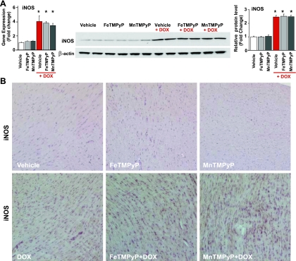
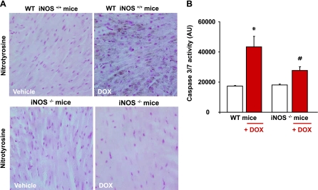
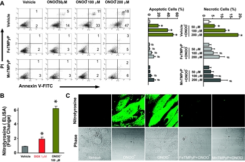
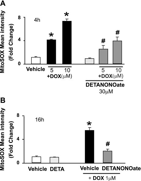
Doxorubicin (DOX) is a potent available antitumor agent; however, its clinical use is limited because of its cardiotoxicity. Cell death is a key component in DOX-induced cardiotoxicity, but its mechanisms are elusive. Here, we explore the role of superoxide, nitric oxide (NO), and peroxynitrite in DOX-induced cell death using both in vivo and in vitro models of cardiotoxicity. Western blot analysis, real-time PCR, immunohistochemistry, flow cytometry, fluorescent microscopy, and biochemical assays were used to determine the markers of apoptosis/necrosis and sources of NO and superoxide and their production. Left ventricular function was measured by a pressure-volume system. We demonstrated increases in myocardial apoptosis (caspase-3 cleavage/activity, cytochrome c release, and TUNEL), inducible NO synthase (iNOS) expression, mitochondrial superoxide generation, 3-nitrotyrosine (NT) formation, matrix metalloproteinase (MMP)-2/MMP-9 gene expression, poly(ADP-ribose) polymerase activation [without major changes in NAD(P)H oxidase isoform 1, NAD(P)H oxidase isoform 2, p22(phox), p40(phox), p47(phox), p67(phox), xanthine oxidase, endothelial NOS, and neuronal NOS expression] and decreases in myocardial contractility, catalase, and glutathione peroxidase activities 5 days after DOX treatment to mice. All these effects of DOX were markedly attenuated by peroxynitrite scavengers. Doxorubicin dose dependently increased mitochondrial superoxide and NT generation and apoptosis/necrosis in cardiac-derived H9c2 cells. DOX- or peroxynitrite-induced apoptosis/necrosis positively correlated with intracellular NT formation and could be abolished by peroxynitrite scavengers. DOX-induced cell death and NT formation were also attenuated by selective iNOS inhibitors or in iNOS knockout mice. Various NO donors when coadministered with DOX but not alone dramatically enhanced DOX-induced cell death with concomitant increased NT formation. DOX-induced cell death was also attenuated by cell-permeable SOD but not by cell-permeable catalase, the xanthine oxidase inhibitor allopurinol, or the NADPH oxidase inhibitors apocynine or diphenylene iodonium. Thus, peroxynitrite is a major trigger of DOX-induced cell death both in vivo and in vivo, and the modulation of the pathways leading to its generation or its effective neutralization can be of significant therapeutic benefit.


Figures

Fig. 1.
Fig. 1.
Effects of doxorubicin (DOX) with or without peroxynitrite scavengers (PSs) on myocardial inducible nitric oxide (NO) synthase (iNOS) expression in vivo. A: DOX-induced increased myocardial iNOS mRNA (left) and protein (right) levels, which were not affected by PSs. *P < 0.05 vs. vehicle; #P < 0.05 vs. DOX. n = 6 per group for protein samples and n = 9 per group for mRNA samples. B: immunohistochemistry demonstrated widespread DOX-induced increased myocardial iNOS expression. FeTMPyP, iron α,β,γ,δ-tetrakis(4-N-methylpyridyl)porphine; MnTMPyP, mangenese α,β,γ,δ-tetrakis(4-N-methylpyridyl)porphine.
Fig. 2.
Fig. 2.
Effects of DOX with or without PSs on myocardial superoxide/ROS formation, its sources, and antioxidant enzyme activities in vivo. A: effects of PSs on DOX-induced superoxide/ROS formation from frozen heart sections using dihydroethidium (DHE). *P < 0.05 vs. vehicle; #P < 0.05 vs. DOX. n = 6 per group. B: effects of PSs on DOX-induced myocardial xanthine oxidase mRNA (left) and protein (right) expression. A representative blot from 3 sets of experiments is shown. The blot was also probed for β-actin as a loading control. *P < 0.05 vs. vehicle; #P < 0.05 vs. DOX. n = 6 per group. C: effects of DOX with or without PSs on mitochondrial superoxide/ROS generation in isolated mitochondria. *P < 0.05 vs. vehicle; #P < 0.05 vs. DOX. n = 6 per group. D: effects of DOX with or without PSs on myocardial SOD activity. n = 6 per group. E and F: effects of DOX with or without PSs on myocardial catalase (E) and glutathione peroxidase (F) activities. *P < 0.05 vs. vehicle; #P < 0.05 vs. DOX. n = 6 per group.
Fig. 3.
Fig. 3.
Effects of DOX with or without PSs on myocardial nitrotyrosine (NT) formation, matrix metalloproteinase (MMP)-2 and MMP-9 gene expression, and poly(ADP-ribose) polymerase (PARP) and myeloperoxidase (MPO) activities in vivo. A: effects of PSs on DOX-induced NT formation from heart tissue homogenates. B: effects of PSs on DOX-induced myocardial MMP-2 and MMP-9 mRNA expression. *P < 0.05 vs. vehicle; #P < 0.05 vs. DOX. n = 9 per group. C: effects of PSs on DOX-induced myocardial PARP activity. *P < 0.05 vs. vehicle; #P < 0.05 vs. DOX. n = 6 per group. D: effects of DOX with or without PSs on myocardial MPO staining. A representative sample from liver ischemia-reperfusion (I/R) injury with marked neutrophil infiltration as previously described (4, 52) and stained under the same conditions was used as a positive control.
Fig. 4.
Fig. 4.
Effects of DOX with or without PSs on myocardial apoptosis in vivo. A: effects of PSs on DOX-induced myocardial cytochrome c (Cyt-C) release from mitochondria. *P < 0.05 vs. vehicle; #P < 0.05 vs. DOX. n = 6 per group. B: effects of PSs on DOX-induced myocardial caspase-3 activation. *P < 0.05 vs. vehicle; #P < 0.05 vs. DOX. n = 6 per group. C: effects of PSs on DOX-induced caspase-3 activity. *P < 0.05 vs. vehicle; #P < 0.05 vs. DOX. n = 6 per group. D: effects of PSs on DOX-induced myocardial DNA fragmentation (TUNEL assay). Eu, europium; AU, arbitrary units. *P < 0.05 vs. vehicle; #P < 0.05 vs. DOX. n = 6 per group. E: effects of PSs on DOX-induced caspase-3 and caspase-9 gene expression. Data were analyzed using two housekeeping genes, and the data presented here were normalized to β-actin. *P < 0.05 vs. vehicle; #P < 0.05 vs. DOX. n = 9 per group.
Fig. 5.
Fig. 5.
Effects of DOX on myocardial NT generation and caspase-3/7 activity in iNOS knockout mice. A and B: decreased DOX-induced NT formation (A) and apoptosis (B) as measured by myocardial caspase-3/7 activity in iNOS−/− mice compared with wild-type (WT) mice. *P < 0.05 vs. vehicle; #P < 0.05 vs. DOX. n = 9 per group.
Fig. 6.
Fig. 6.
Effects of DOX with or without PSs on myocardial function in vivo. A: effect of PSs on the DOX-induced depression of left ventricular systolic pressure (LVSP), maximal slope of the systolic pressure increment (+dP/dt), ejection fraction, stroke work (SW), and cardiac output in mice. B: effects of PSs on the DOX-induced depression of load-independent indexes of cardiac contractility [dP/dt-end-diastolic volume (EDV) relation, preload-recruitable SW (PRSW), and maximal elastance (Emax)]. Hemodynamic parameters were measured 5 days after DOX administration. Results are means ± SE of 6–12 experiments in each group. *P < 0.05 vs. vehicle; #P < 0.05 vs. DOX.
Fig. 7.
Fig. 7.
Effects of DOX with or without PSs and/or iNOS inhibitors on mitochondrial superoxide generation and cell death in H9c2 cardiomyocytes in vitro. A: effects of DOX with or without PSs on cell death and mitochondrial superoxide generation in vitro as measured by quantitative flow cytometry. *P < 0.05 vs. vehicle; #P < 0.05 vs. DOX. n = 6 per group. B: effects of iNOS inhibitors {S,S′-[1,3-phenylene-bis(1,2-ethanediyl)]bis-isothiourea (1,3-PB-ITU) and l-N6-(1-iminorthyl)-lysine (l-NIL)} on DOX-induced cell death in vitro. *P < 0.05 vs. vehicle; #P < 0.05 vs. DOX. n = 9 per group.
Fig. 8.
Fig. 8.
Effects of PSs on DOX-induced mitochondrial dysfunction in vitro. A: effects of PSs (200 μM) on DOX-induced dissipation of mitochondrial membrane potential (as measured by TMRE) in H9c2 cells using flow cytometry. Various controls are also shown at the far right, including vehicle without TMRE, vehicle with TMRE, and dissipation of mitochondrial potential by FCCP + oligomycin A (Oligo A) for 2 and 30 min after exposure. B: effects of PSs (200 μM) on DOX-induced dissipation of mitochondrial membrane potential measured in H9c2 cells loaded with JC-1 using confocal microscopy. Magnification: ×600.
Fig. 9.
Fig. 9.
Effects of PSs on DOX-induced apoptosis/necrosis in vitro. A: effects of PSs (200 μM) on DOX-induced apoptosis/necrosis as measured by flow cytometry in H9c2 cardiomyocytes. Representative data from 6 separate experiments were analyzed. PI, propidium iodide. *P < 0.05 vs. vehicle; #P < 0.05 vs. DOX. n = 6 per group. B: effects of PSs on DOX-induced Cyt-C release as analyzed by Western blot from H9c2 cells. Shown is a representative blot from 3 separate experiments. *P < 0.05 vs. vehicle; #P < 0.05 vs. DOX. n = 6 per group. C: effects of PSs on DOX-induced active caspase expression (green) and the nuclear staining pattern by Hoechst 33342 dye (blue). Representative data from 10 experiments were analyzed. Magnification: ×150.
Fig. 10.
Fig. 10.
Effects of PSs on peroxynitrite-induced apoptosis/necrosis and NT generation in vitro. A: effects of PSs (200 μM) on peroxynitrite-induced apoptosis/necrosis as measured by flow cytometry in H9c2 cardiomyocytes. The treatment of peroxynitrite was described previously (Ref. 22). Representative data from 4 experiments were analyzed. *P < 0.05 vs. vehicle; #P < 0.05 vs. DOX. n = 4 per group. B: effects of DOX and peroxynitrite on NT formation in H9c2 cells as measured by quantitative ELISA. *P < 0.05 vs. vehicle; n = 4 per group. C: effects of PSs on peroxynitrite-induced NT formation (green). Representative data from 8 experiments are shown. Magnification: ×600.
Fig. 11.
Fig. 11.
Effects of DOX with or without PSs on intracellular NT formation. Shown are the effects of PSs on the intracellular localization of DOX-induced NT formation (red) and the mitochondrial dye Mitotracker green by confocal microscopy. Magnification: ×800.
Fig. 12.
Fig. 12.
Effects of the NO donor diethylenetriamine (DETA) NONOate on DOX-induced apoptosis/necrosis in vitro. A: effects of the NO donor DETA NONOate (30 and 100 μM) on DOX-induced apoptosis/necrosis as measured by flow cytometry in H9c2 cardiomyocytes. Representative data from 4 experiments were analyzed. B: percentages of apoptotic (top) and necrotic (bottom) cells. *P < 0.05 vs. vehicle; #P < 0.05 vs. DOX. n = 4 per group.
Fig. 13.
Fig. 13.
Effect of DOX with or without the NO donor DETA NONOate on cell death and intracellular NT generation in vitro. A: DOX dose dependently increased apoptosis and necrosis in H9c2 cardiomyocytes (left), which were drastically enhanced by the addition of the NO donor DETA NONOate (right). DETA NONOate by itself only slightly increased apoptosis but not necrosis at high concentrations. Similar results were observed with 2 additional NO donors (see supplemental Fig. 7). B: DOX dose dependently increased intracellular NT formation in H9c2 cardiomyocytes (left), which was enhanced by the addition of the NO donor DETA NONOate (left and right). DETANONOate by itself did not increase intracellular NT formation in H9c2 cardiomyocytes. C: correlation between intracellular NT formation (as measured by ELISA) and apoptosis/necrosis in H9c2 cardiomyocytes. The correlation analysis included vehicle-, DOX-, and DOX + DETA NONOate-treated cells. Each point in the graph represents a flow cytometry experiment.
Fig. 14.
Fig. 14.
Effects of NO donors on DOX-induced mitochondrial superoxide formation in vitro. Shown are the effects of NO donors in both short-term (4 h; A) and long-term (16 h; B) DOX-induced mitochondrial superoxide formation. *P < 0.001 vs. vehicle; #P < 0.001 vs. the corresponding DOX concentration. n = 6 per group.
Fig. 15.
Fig. 15.
Schematic diagram of DOX-induced cardiotoxicity: role of superoxide, NO, and peroxynitrite. DOX initially increases mitochondrial superoxide and, consequently, the generation of other ROS (e.g., H2O2) in cardiomyocytes and/or endothelial cells by redox cycling. Increased DOX-induced ROS generation in cardiomyocytes triggers the activation of the transcription factor NF-κB, leading to enhanced iNOS expression and NO generation. NO reacts with superoxide to form peroxynitrite both in the cytosol and mitochondria, which, in turn, induces cell damage via lipid peroxidation, inactivation of enzymes and other proteins by oxidation and nitration, and activation of stress signaling pathways (e.g., MAPK), MMPs, and PARP-1, among others. In the mitochondria, peroxynitrite, in concert with other ROS/reactive nitrogen species, impairs various key mitochondrial enzymes, leading to more sustained intracellular ROS generation (persistent even after DOX already metabolized), triggering further activation of transcription factor(s) and iNOS expression, resulting in the amplification of oxidative/nitrosative stress. In the mitochondria, peroxynitrite also triggers the release of proapoptotic factors (e.g., Cyt-C and apoptosis-inducing factor) mediating caspase-dependent and -independent cell death pathways, which are also pivotal in DOX-induced cardiotoxicity. Peroxynitrite, in concert with other oxidants, also causes strand breaks in DNA, activating the nuclear enzyme PARP-1. Once excessive oxidative and nitrosative stress-induced DNA damage occurs, overactivated PARP initiates an energy-consuming cycle by transferring ADP-ribose units from NAD+ to nuclear proteins, resulting in the rapid depletion of intracellular NAD+ and ATP pools, slowing the rate of glycolysis and mitochondrial respiration, eventually leading to cellular dysfunction and death, mostly by necrosis. Overactivated PARP may also facilitate the expression of a variety of inflammatory genes leading to increased inflammation (PARP-1 is a known coactivator of NF-κB) and associated oxidative stress, thus facilitating the progression of cardiovascular dysfunction and heart failure. PARG, poly(ADP-ribose) glycohydrolase.

References

    1. Altmann SM, Muryshev A, Fossale E, Maxwell MM, Norflus FN, Fox J, Hersch SM, Young AB, MacDonald ME, Abagyan R, Kazantsev AG. Discovery of bioactive small-molecule inhibitor of poly ADP-ribose polymerase: implications for energy-deficient cells. Chem Biol 13: 765–770, 2006. - PubMed
    1. Andreadou I, Sigala F, Iliodromitis EK, Papaefthimiou M, Sigalas C, Aligiannis N, Savvari P, Gorgoulis V, Papalabros E, Kremastinos DT. Acute doxorubicin cardiotoxicity is successfully treated with the phytochemical oleuropein through suppression of oxidative and nitrosative stress. J Mol Cell Cardiol 42: 549–558, 2007. - PubMed
    1. Bai P, Mabley JG, Liaudet L, Virág L, Szabó C, Pacher P. Matrix metalloproteinase activation is an early event in doxorubicin-induced cardiotoxicity. Oncol Rep 11: 505–508, 2004. - PubMed
    1. Bátkai S, Osei-Hyiaman D, Pan H, El-Assal O, Rajesh M, Mukhopadhyay P, Hong F, Harvey-White J, Jafri A, Haskó G, Huffman JW, Gao B, Kunos G, Pacher P. Cannabinoid-2 receptor mediates protection against hepatic ischemia/reperfusion injury. FASEB J 21: 1788–1800, 2007. - PMC - PubMed
    1. Bátkai S, Rajesh M, Mukhopadhyay P, Haskó G, Liaudet L, Cravatt BF, Csiszár A, Ungvári Z, Pacher P. Decreased age-related cardiac dysfunction, myocardial nitrative stress, inflammatory gene expression, and apoptosis in mice lacking fatty acid amide hydrolase. Am J Physiol Heart Circ Physiol 293: H909–H918, 2007. - PMC - PubMed

Publication types

MeSH terms