

doi: 10.1074/jbc.M900571200.
Epub 2009 Mar 13.
A structural model for the damage-sensing complex in bacterial nucleotide excision repair
Affiliations
- PMID: 19287003
- PMCID: PMC2676014
- DOI: 10.1074/jbc.M900571200
Item in Clipboard
A structural model for the damage-sensing complex in bacterial nucleotide excision repair
J Biol Chem.
.
Abstract
Nucleotide excision repair is distinguished from other DNA repair pathways by its ability to process a wide range of structurally unrelated DNA lesions. In bacteria, damage recognition is achieved by the UvrA.UvrB ensemble. Here, we report the structure of the complex between the interaction domains of UvrA and UvrB. These domains are necessary and sufficient for full-length UvrA and UvrB to associate and thereby form the DNA damage-sensing complex of bacterial nucleotide excision repair. The crystal structure and accompanying biochemical analyses suggest a model for the complete damage-sensing complex.
Figures
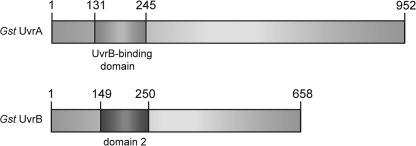
The location of the interaction domains in the primary sequence of
G. stearothermophilus UvrA and UvrB.
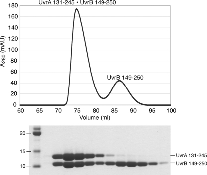
Purification of the UvrA·UvrB interaction domain complex for
structural studies. The complex was formed by mixing UvrA 131–245
with molar excess of UvrB 149–250 and purified by size exclusion
chromatography.
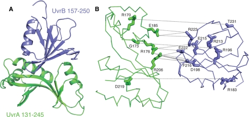
Structural basis for UvrA-UvrB interaction. UvrA 131–245 and
UvrB 157–250 are shown in green and blue,
respectively. A, overall structure of the complex between the
interaction domains of UvrA (residues 131–245) and UvrB (residues
157–250) shown as a ribbon diagram. B, exploded view of the
interaction interface. The interface is largely polar, consisting of a large
number of direct and water-mediated hydrogen bonds, as well as electrostatic
interactions between conserved residues. UvrA and UvrB interaction domains are
shown as Cα trace. Residues that are involved in direct
contacts across the interface are shown as sticks. The interactions
are drawn as dashed lines. This view was generated by separating the
two proteins by 10 Å and rotating them by 65° away from each other.
This orientation was chosen to most clearly depict the interactions (see
Table 2 for a complete list of
interactions).
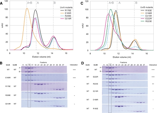
Site-directed mutagenesis in the context of full-length proteins
confirmed the importance of the observed interactions. Interaction between
UvrA and UvrB (8 nmol each) was analyzed by size exclusion chromatography
(Superdex 200; GE Healthcare) at 4 °C in UvrAB complex buffer (20
mm Tris-HCl, pH 7.5, 150 mm KCl, 5% (v/v) glycerol, 5
mm β-ME, 5 mm MgCl2, 2 mm
ATP). A and B, elution profile and SDS-PAGE analysis of the
samples containing mutant UvrA and wild-type UvrB. C and D,
elution profile and SDS-PAGE analysis of the samples containing wild-type UvrA
and mutant UvrB. Disruption of UvrA-UvrB interaction can be clearly seen in
fraction 15 (gray box) in C and D.
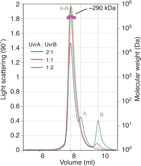
Multi-angle laser light scattering suggests a 2:1 stoichiometry for the
full-length UvrA·UvrB complex. UvrA·UvrB complex was formed
using different UvrA:UvrB ratios and subjected to size exclusion
chromatography (20 mm Tris-HCl, pH 7.5, 150 mm KCl, 5%
(v/v) glycerol, 5 mm β-ME, 5 mm MgCl2, 2
mm ATP), and multi-angle laser light scattering. The complex
appeared monodisperse with an apparent molecular mass of ∼290 kDa,
approximating that of UvrA2·UvrB1.
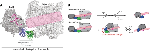
Model of the nucleotide excision repair damage sensor. A,
modeled UvrA·UvrB complex based on superposition of the corresponding
domains onto the experimentally determined structure. Note that the proposed
DNA-binding path on UvrA (5)
and the DNA-binding site on UvrB (Protein Data Bank code 2FDC
(25)) are aligned
(pink). B, the models for lesion recognition by the
UvrA·UvrB complex.
References
-
- Truglio, J. J., Croteau, D. L., Van Houten, B., and Kisker, C. (2006) Chem. Rev. 106 233-252 - PubMed
-
- Lin, J. J., and Sancar, A. (1992) J. Biol. Chem. 267 17688-17692 - PubMed
-
- Verhoeven, E. E., van Kesteren, M., Moolenaar, G. F., Visse, R., and Goosen, N. (2000) J. Biol. Chem. 275 5120-5123 - PubMed
-
- Orren, D. K., Selby, C. P., Hearst, J. E., and Sancar, A. (1992) J. Biol. Chem. 267 780-788 - PubMed
Publication types
MeSH terms
Substances
Associated data
- Actions
Grants and funding
LinkOut - more resources
Full Text Sources
