. 2009 May 15;284(20):13924-13939.
doi: 10.1074/jbc.M901017200. Epub 2009 Mar 14.

Clathrin regulates the association of PIPKIgamma661 with the AP-2 adaptor beta2 appendage

Affiliations

Clathrin regulates the association of PIPKIgamma661 with the AP-2 adaptor beta2 appendage

James R Thieman et al. J Biol Chem. .

Abstract

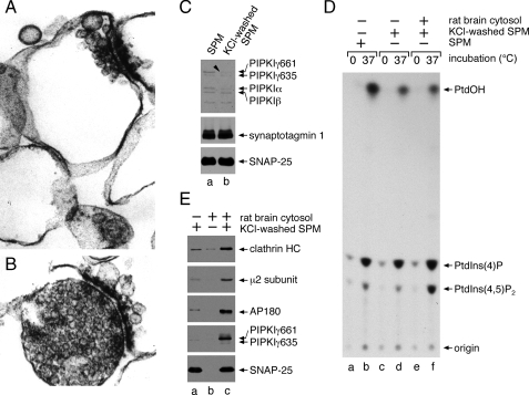
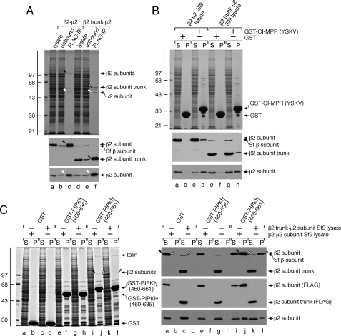
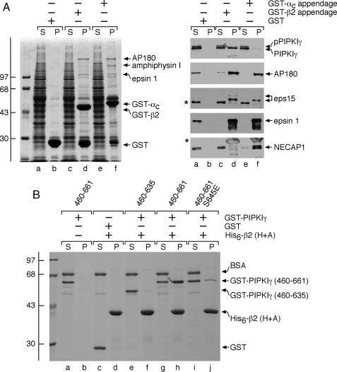
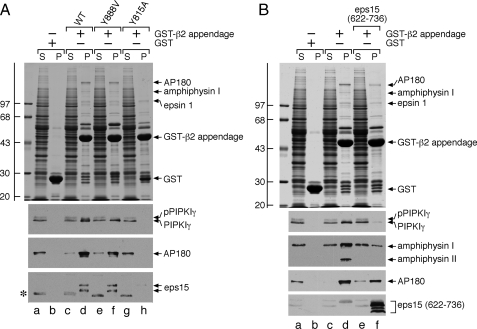
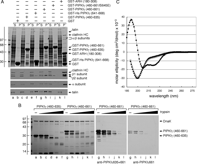
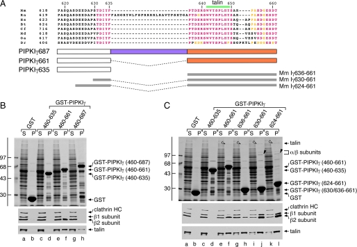
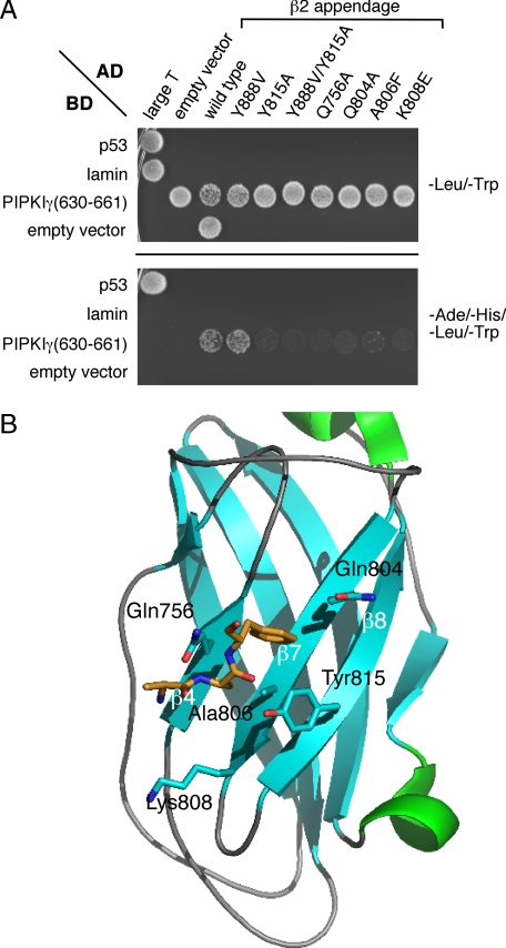
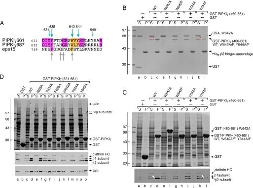
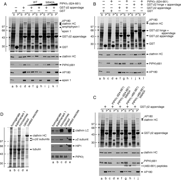
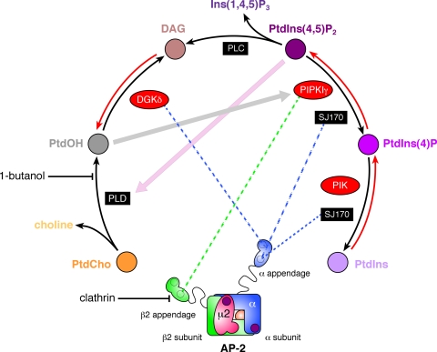
The AP-2 clathrin adaptor differs fundamentally from the related AP-1, AP-3, and AP-4 sorting complexes because membrane deposition does not depend directly on an Arf family GTPase. Instead phosphatidylinositol 4,5-bisphosphate (PtdIns(4,5)P(2)) appears to act as the principal compartmental cue for AP-2 placement at the plasma membrane as well as for the docking of numerous other important clathrin coat components at the nascent bud site. This PtdIns(4,5)P(2) dependence makes type I phosphatidylinositol 4-phosphate 5-kinases (PIPKIs) lynchpin enzymes in the assembly of clathrin-coated structures at the cell surface. PIPKIgamma is the chief 5-kinase at nerve terminals, and here we show that the 26-amino acid, alternatively spliced C terminus of PIPKIgamma661 is an intrinsically unstructured polypeptide that binds directly to the sandwich subdomain of the AP-2 beta2 subunit appendage. An aromatic side chain-based, extended interaction motif that also includes the two bulky C-terminal residues of the short PIPKIgamma635 variant is necessary for beta2 appendage engagement. The clathrin heavy chain accesses the same contact surface on the AP-2 beta2 appendage, but because of additional clathrin binding sites located within the unstructured hinge segment of the beta2 subunit, clathrin binds the beta2 chain with a higher apparent affinity than PIPKIgamma661. A clathrin-regulated interaction with AP-2 could allow PIPKIgamma661 to be strategically positioned for regional PtdIns(4,5)P(2) generation during clathrin-coated vesicle assembly at the synapse.


Figures

FIGURE 1.
FIGURE 1.
PIPKIγ661 and AP-2 are coordinately recruited to the synaptic plasma membrane. A and B, aliquots of synaptic plasma membrane (A) or synaptosomes from which they were derived (B) were fixed with 2% glutaraldehyde and processed for electron microscopy. Thin section micrographs typical of the many fields examined are shown. C, samples of 50 μg of synaptic plasma membrane (SPM), suspended in assay buffer alone or supplemented with 1 m KCl, were sedimented after incubation on ice for 30 min. Aliquots of each resuspended membrane pellet were prepared for SDS-PAGE and immunoblotting. Portions of the blots were probed with an affinity-purified polyclonal anti-PIPKI antibody or mAbs directed against synaptotagmin 1 or SNAP-25. D, reactions containing 0.5 mg/ml untreated or salt-washed synaptic plasma membranes, 5 mg/ml cytosol, and 500 μm [γ-32P]ATP were prepared as indicated. After incubation at 37 °C for 10 min the lipids were extracted and analyzed by TLC and autoradiography. A representative experiment of three is shown, and the migration positions of authentic phospholipid standards are indicated. E, reactions containing 50 μg/ml salt-washed synaptic plasma membranes, 5 mg/ml rat brain cytosol, and an ATP-regenerating system were prepared as indicated. After incubation at 37 °C for 15 min, membranes were sedimented and prepared for SDS-PAGE and immunoblotting. Portions of the blots were probed with anti-clathrin subunit heavy chain (HC) mAb TD.1, rabbit R11-29 anti-μ2 serum, or an anti-AP180, anti-PIPKIγ, or anti-SNAP-25 mAb, and only relevant portions of the blots are shown. PtdIns(4)P, phosphatidylinositol 4-phosphate.
FIGURE 2.
FIGURE 2.
The C terminus of PIPKIγ661 but not PIPKIγ635 binds selectively to the AP-2 β2 appendage. A, equivalent volumes of Sf9 cell lysate overexpressing either the β2·μ2 or the β2 trunk·μ2 hemicomplex before (lanes a and d) or after (lanes b and e) immunoprecipitation with agarose-coupled anti-FLAG mAb M2 and aliquots from the washed and FLAG peptide-eluted immunoprecipitate (IP) pellet (lanes c and f) were resolved by SDS-PAGE and either stained with Coomassie Blue or transferred to nitrocellulose. Portions of the blots were probed with anti-β1/β1-subunit mAb 100/1 or anti-μ2 serum, and only the relevant portions are shown. The migration position of the molecular mass standards is indicated on the left, and the location of the immunoprecipitated β2 subunit (black arrowhead), β2 trunk (open arrowhead), and μ2 subunit (white arrowheads) are shown. B, ∼250 μg of GST (lanes a, b, e, and f) or GST-CI-MPR (YSKV; lanes c, d, g, and h) immobilized on glutathione-Sepharose was incubated with Sf9 cell lysates overexpressing either the β2·μ2(lanes a-d) or β2 trunk·μ2(lanes e-h) hemicomplexes as indicated. After centrifugation, aliquots of ∼1.5% of each supernatant (S) and ∼12.5% of each washed pellet (P) were resolved by SDS-PAGE and either stained with Coomassie Blue or transferred to nitrocellulose. Portions of the blots were probed with anti-β1/β1-subunit mAb 100/1 or anti-μ2 serum, and only the relevant portions are shown. C, ∼200 μg of GST (lanes a-d), GST-PIPKIγ-(460-635) (lanes e-h), or GST-PIPKIγ-(460-661) immobilized on glutathione beads was incubated with Sf9 cell lysates overexpressing either the β2·μ2(lanes a, b, e, f, i, and j) or β2 trunk·μ2(lanes c, d, g, h, k, and l) hemicomplexes. After centrifugation, aliquots of ∼1.5% of each supernatant (S) and ∼15% of each washed pellet (P) were resolved by SDS-PAGE and either stained with Coomassie Blue or transferred to nitrocellulose. Portions of the blots were probed with anti-β1/β1-subunit mAb 100/1, anti-FLAG mAb M1, or anti-μ2 serum, and only the relevant portions are shown. The migration positions of the FLAG-tagged β2(arrowheads) and the presumptive Sf9 cell β subunit (open arrowheads) are indicated.
FIGURE 3.
FIGURE 3.
PIPKIγ661 discriminates between the AP-2α andβ2 appendages. A, ∼100 μg of GST (lanes a and b), GST-β2 appendage (lanes c and d), or GST-αC appendage (lanes e and f) immobilized on glutathione-Sepharose was incubated with rat brain cytosol as indicated. After centrifugation, aliquots of ∼1.5% of each supernatant (S) and ∼10% of each washed pellet (P) were resolved by SDS-PAGE and either stained with Coomassie Blue or transferred to nitrocellulose. Portions of the blots were probed with anti-PIPKIγ mAb clone 12, anti-AP180 mAb clone 34, or affinity-purified anti-eps15, -epsin 1, or -NECAP 1 antibodies, and only the relevant portions are shown. The different migration of the non-phosphorylated (arrowhead) and phosphorylated (pPIPKIγ) forms of PIPKIγ is indicated. The asterisk demarcates a non-specific band detected by the anti-eps15 antibodies. B, ∼5 μg of His6-tagged β2 hinge + appendage (H+A; lanes c-j) immobilized on Ni-NTA-agarose was incubated with ∼25 μg of GST (lanes c and d), GST-PIPKIγ-(460-635) (lanes e and f), GST-PIPKIγ-(460-661) (lanes g and h), or GST-PIPKIγ-(460-661) with a phosphomimetic S645E mutation (lanes i and j) as indicated in the presence of carrier bovine serum albumin (BSA). GST-PIPKIγ-(460-661) was also incubated with Ni-NTA-agarose alone (lanes a and b). After centrifugation, aliquots of ∼2.5% of each supernatant (S) and ∼25% of each washed pellet (P) were resolved by SDS-PAGE and stained with Coomassie Blue.
FIGURE 4.
FIGURE 4.
PIPKIγ661 physically contacts the sandwich subdomain of the AP-2 β2 appendage. A, ∼100 μg of GST (lanes a and b), GST-β2 appendage (lanes c and d), or GST-β2 Y888V (lanes e and f) or Y815A (lanes g and h) mutant appendage immobilized on glutathione-Sepharose was incubated with rat brain cytosol as indicated. After centrifugation, aliquots of ∼1.5% of each supernatant (S) and ∼10% of each washed pellet (P) were resolved by SDS-PAGE and either stained with Coomassie Blue or transferred to nitrocellulose. Portions of the blots were probed with anti-PIPKIγ mAb clone 12, anti-AP180 mAb clone 34, or affinity-purified anti-eps15 antibodies, and only the relevant portions are shown. The slowed migration of the phosphorylated from of PIPKIγ (pPIPKIγ) is indicated as is the location of the Coomassie Blue-stained epsin 1 band. The asterisk demarcates a non-specific band detected by the anti-eps15 antibodies. B, ∼100 μg of GST (lanes a and b) or GST-β2 appendage (lanes c-f) immobilized on glutathione-Sepharose was incubated with rat brain cytosol in the absence or presence of 2 μm eps15 (residues 622-736) competitor polypeptide (lanes e and f) as indicated. After centrifugation, aliquots of ∼1.5% of each supernatant (S) and ∼10% of each washed pellet (P) were resolved by SDS-PAGE and either stained with Coomassie Blue or transferred to nitrocellulose. Portions of the blots were probed with anti-PIPKIγ mAb clone 12, anti-amphiphysin mAb clone 15, anti-AP180 mAb clone 34, or anti-eps15 antibodies, and only the relevant portions are shown. The slowed migration of the phosphorylated from of PIPKIγ (pPIPKIγ) is indicated, as is the location of the Coomassie Blue-stained epsin 1 band. WT, wild type.
FIGURE 5.
FIGURE 5.
The alternatively spliced C terminus of PIPKIγ alone does not contain all the AP-2 binding information. A, ∼200 μg of GST (lanes a and b), GST-PIPKIγ-(460-635) (lanes c and d), GST-Hs PIPKIγ-(641-668) (lanes e and f), GST-PIPKIγ-(460-661) (lanes g and h), GST-PIPKIγ-(460-661) containing S645E (lanes i and j), or GST-ARH-(180-308) (lanes k and l) immobilized on glutathione-Sepharose was incubated with rat brain cytosol as indicated. After centrifugation, aliquots of ∼1.5% of each supernatant (S) and ∼10% of each washed pellet (P) were resolved by SDS-PAGE and either stained with Coomassie Blue or transferred to nitrocellulose. Portions of the blots were probed with anti-clathrin heavy chain (HC) mAb TD.1 and anti-β1/β2 subunit mAb 100/1, anti-AP-2 α subunit mAb C4, or anti-talin mAb 8d4, and only the relevant portions are shown. The position of talin (open arrowheads) and the AP-2 β2 and α subunits (black arrowheads) on the stained gel is shown. Note that PIPKIγ661 clearly binds to the β2 appendage of AP-2 more weakly than ARH, which has a KD for AP-2 of ∼2 μm (49, 50). B, aliquots (20 μg) of PIPKIγ-(460-635) (lanes a-f) or PIPKIγ-(460-661) (lanes g-l) were incubated alone or with 1:5 serial 5-fold dilutions of trypsin (0.6-375 ng) in 50 mm Tris, pH 8.0, 150 mm NaCl, 5 mm CaCl2, 1 mm DTT at 37 °C for 1 h. Proteolysis was stopped by the addition of 95 °C SDS sample buffer, and aliquots of 10% of each reaction were resolved by SDS-PAGE and either stained with Coomassie Blue or transferred to nitrocellulose. The PIPKIγ-(460-661) portion of the blot was probed with anti-PIPKIγ mAb clone 12 that recognizes both isoforms or with an affinity-purified antibody that recognizes only the PIPKIγ661 isoform. C, circular dichroism spectra of PIPKIγ-(460-635) (open circles), PIPKIγ-(460-661) (triangles), and epsin 1 ENTH domain (squares) polypeptides were measured in 25 mm potassium phosphate + 1 mm DTT. Measurements were made from 280 to 185 nm at 1-nm increments, and the spectra were base line-corrected and represent the average of five (PIPKIγ) or three (ENTH) runs. Note the signature α-helical features of the globular ENTH domain compared with the unstructured PIPKIγ polypeptides. deg, degrees.
FIGURE 6.
FIGURE 6.
Differing partner binding properties of the three PIPKIγ splice variants. A, primary sequence alignment of the C-terminal segment of PIPKIγ isoforms from various species: murine (Mm; NCBI accession number NP_032870.1), rat (Rn; NP_001009967.2), human (Hs; NP_036530.1), bovine (Bt; XP_585653.4), feline (Cf; XP_542172.2), opossum (Md; XP_001363745.1), platypus (Oa; XP_001511349.1), and zebrafish (Dr; XP_683392.3). Identical residues are colored pink, and conservatively substituted residues are yellow. The location of the talin-binding motif is indicated above, and the GST fusion proteins spanning the junction between the PIPKIγ635 and -661 isoforms tested is indicated below. B, ∼200 μg of GST (lanes a and b), GST-PIPKIγ-(460-635) (lanes c and d), GST-PIPKIγ-(460-661) (lanes e and f), or GST-PIPKIγ-(460-687) (lanes g and h) immobilized on glutathione-Sepharose was incubated with rat brain cytosol as indicated. After centrifugation, aliquots of 1.5% of each supernatant (S) and 10% of each washed pellet (P) were resolved by SDS-PAGE and either stained with Coomassie Blue or transferred to nitrocellulose. Portions of the blots were probed with anti-clathrin heavy chain (HC) mAb TD.1 and anti-β1/β2 subunit mAb 100/1 or with anti-talin mAb 8d4, and only the relevant portions are shown. C, ∼200 μg of GST (lanes a and b), GST-PIPKIγ-(460-635) (lanes c and d), GST-PIPKIγ-(460-661) (lanes e and f), GST-PIPKIγ-(636-661) (lanes g and h), GST-PIPKIγ-(630-661) (lanes i and j), or GST-PIPKIγ-(624-661) (lanes k and l) immobilized on glutathione-Sepharose was incubated with rat brain cytosol as indicated. After centrifugation, aliquots of 1.5% of each supernatant(S) and 10% of each washed pellet (P) were resolved by SDS-PAGE and either stained with Coomassie Blue or transferred to nitrocellulose. Portions of the blots were probed with anti-clathrin heavy chain (HC) mAb TD.1 and anti-β1/β2 subunit mAb 100/1 or anti-talin mAb 8d4, and only the relevant portions are shown. The position of talin (open arrowheads) and the AP-2 β2 and αC subunits (black arrowheads) on the stained gel is shown.
FIGURE 7.
FIGURE 7.
AP-2-PIPKIγ interaction in a yeast two-hybrid assay. A, S. cerevisiae strain AH109 transformed with the indicated Gal4 pGBKT7 binding domain (BD) and pGADT7 activation domain (AD) plasmid combinations were spotted onto synthetic defined minimal medium plates lacking either Leu and Trp or Ade, His, Leu, and Trp and grown at 30 °C. B, ribbon representation (Protein Data Bank code 2G30) of the AP-2 β2 appendage sandwich subdomain indicating the location of important side chains (blue, nitrogen; red, oxygen) involved in accommodating the PIPKIγ661 C-terminal interaction motif. Shown in stick representation (gold) is the location of co-crystallizing AAF peptide that demarcates a portion of the binding surface upon the β2 appendage sandwich subdomain.
FIGURE 8.
FIGURE 8.
Delineation of key anchor residues that mediate PIPKIγ661 binding to the β2 appendage. A, primary sequence alignment of the two PIPKIγ 26-amino acid inserts and comparison with a tract from eps15 that also binds to the AP-2 β2 appendage sandwich subdomain. B, ∼40 μg of His6-tagged β2 hinge + appendage immobilized on Ni-NTA-agarose was incubated with ∼10 μg of GST (lanes a and b), GST-PIPKIγ-(460-661) wild type (WT; lanes c and d), or GST-PIPKIγ-(460-661) containing W642A (lanes e and f), W642F (lanes g and h), Y644A (lanes i and j), or Y644F (lanes k and l) mutations in the presence of carrier bovine serum albumin (BSA). After centrifugation, aliquots of ∼4% of each supernatant (S) and ∼25% of each washed pellet (P) were resolved by SDS-PAGE and stained with Coomassie Blue. Bound protein (red arrowhead) is shown. C, ∼250 μg of GST (lanes a and b), GST-PIPKIγ-(460-661) wild type (WT; lanes c and d), or GST-PIPKIγ-(460-661) containing W642A (lanes e and f), W642F (lanes g and h), Y644A (lanes i and j), or Y644F (lanes k and l) mutations immobilized on glutathione-Sepharose was incubated with rat brain cytosol as indicated. After centrifugation, aliquots of ∼3.5% of each supernatant (S) and ∼10% of each pellet (P) were resolved by SDS-PAGE and either stained with Coomassie Blue or transferred to nitrocellulose. A portion of the blot was probed with anti-β1/β2-subunit mAb 100/1 and anti-clathrin heavy chain (HC) mAb TD.1. Bound protein (white arrowhead) is shown. D, ∼100 μg of GST (lanes a and b), GST-PIPKIγ-(624-661) wild type (WT; lanes c and d), or GST-PIPKIγ-(624-661) containing I633A (lanes e and f), Y634A (lanes g and h), F635A (lanes i and j), W642A (lanes k and l), Y644A (lanes m and n), or Y649A (lanes o and p) mutations immobilized on glutathione-Sepharose was incubated with rat brain cytosol as indicated. After centrifugation, aliquots of ∼1.5 supernatant (S) and ∼10% of each pellet (P) were resolved by SDS-PAGE and either stained with Coomassie Blue or transferred to nitrocellulose. Portions of the blot were probed with anti-β1/β2-subunit mAb 100/1 and anti-clathrin heavy chain (HC) mAb TD.1 or anti-talin mAb 8d4. The position of talin (open arrowhead) and the AP-2 α and β2 subunits (black arrowhead) on the stained gel are shown.
FIGURE 9.
FIGURE 9.
Mutually exclusive engagement of the β2 appendage sandwich by either PIPKIγ661 or clathrin. A, ∼100 μg of GST (lanes a and b) or GST-β2 appendage (lanes c-l) immobilized on glutathione-Sepharose was incubated with rat brain cytosol alone (lanes a-d) or cytosol supplemented with 46 μm wild type (WT; lanes e and f) or W642A (lanes i and j) PIPKIγ-(624-661) peptide or 139 μm wild type (lanes g and h) or W642A (lanes k and l) PIPKIγ-(624-661) peptide. After centrifugation, aliquots of ∼1.5% of each supernatant (S) and ∼10% of each washed pellet (P) were resolved by SDS-PAGE and either stained with Coomassie Blue or transferred to nitrocellulose. Portions of the blots were probed with anti-clathrin heavy chain (HC) mAb TD.1, anti-PIPKIγ mAb clone 12, anti-AP180 mAb clone 34, or affinity-purified anti-epsin 1 antibodies. Notice that although epsin 1 binding to the β2 appendage is affected by the addition of the PIPKIγ peptide, particularly at the highest concentration, the sandwich-binding partners are clearly much more sensitive to the competitor. B, ∼100 μg of GST (lanes a-d), GST-β2 appendage (lanes e-h), or GST-β2 hinge + appendage (lanes i-l) immobilized on glutathione-Sepharose was incubated with rat brain cytosol alone (lanes a, b, e, f, i, and j) or rat brain cytosol supplemented with 113 μm PIPKIγ-(624-661) polypeptide (lanes c, d, g, h, k, and l). After centrifugation, aliquots of ∼1.5% of each supernatant (S) and ∼10% of each washed pellet (P) were resolved by SDS-PAGE and either stained with Coomassie Blue or transferred to nitrocellulose. Portions of the blots were probed with anti-clathrin heavy chain (HC) mAb TD.1, anti-PIPKIγ mAb clone 12, or anti-AP180 mAb clone 34. C, ∼100 μg of GST-β2 appendage immobilized on glutathione-Sepharose was incubated with rat brain cytosol alone (lanes a and b) or cytosol supplemented with 113 μm wild type (lanes c and d) or W642A (lanes e and f) PIPKIγ-(624-661) polypeptide or 113 μm wild type (lanes g and h) or W642A (lanes i and j) PIPKIγ-(460-661) polypeptide. After centrifugation, aliquots of ∼1.5% of each supernatant (S) and ∼10% of each washed pellet (P) were resolved by SDS-PAGE and either stained with Coomassie Blue or transferred to nitrocellulose. Portions of the blots were probed with anti-clathrin heavy chain (HC) mAb TD.1, anti-PIPKIγ mAb clone 12, or anti-AP180 mAb clone 34. D, fractions (20 μg) from a preparation of rat brain clathrin-coated vesicles were resolved by SDS-PAGE and either stained with Coomassie Blue or transferred to nitrocellulose. Portions of the blots were probed with anti-clathrin light chain (LC) mAb Cl57.3, anti-μ2 subunit serum, affinity-purified anti-HIP1 antibodies, or anti-PIPKIγ mAb clone 12. Only the relevant portions are shown. Note the strong enrichment of clathrin, AP-2, and HIP1 (arrowheads) but exclusion of PIPKIγ (open arrowhead) in the coated vesicle fraction.
FIGURE 10.
FIGURE 10.
Phosphoinositide metabolism at clathrin-coated buds. Schematic representation of key enzymes responsible for PtdIns(4,5)P2 synthesis and the mapped interactions with the AP-2 adaptor appendages. The location of the two separate PtdIns(4,5)P2 binding sites on the AP-2 heterotetramer is indicated with purple spots. DAG, diacylglycerol; PLD, phospholipase D; PLC, phospholipase C; PIK, phosphatidylinositol 4-kinase; PtdCho, phosphatidylcholine; Ins(1,4,5)P3, inositol 1,4,5-trisphosphate; PtdIns, phosphatidylinositol; PtdIns(4)P, phosphatidylinositol 4-phosphate.

Similar articles

Cited by

References

    1. Krauss, M., and Haucke, V. (2007) EMBO Rep. 8 241-246 - PMC - PubMed
    1. Di Paolo, G., and De Camilli, P. (2006) Nature 443 651-657 - PubMed
    1. Maldonado-Baez, L., and Wendland, B. (2006) Trends Cell Biol. 16 505-513 - PubMed
    1. Schmid, E. M., and McMahon, H. T. (2007) Nature 448 883-888 - PubMed
    1. Ungewickell, E. J., and Hinrichsen, L. (2007) Curr. Opin. Cell Biol. 19 417-425 - PubMed

Publication types

MeSH terms

Substances