. 2009 Feb 28;41(2):94-101.
doi: 10.3858/emm.2009.41.2.012.

STAT3 is involved in phosphatidic acid-induced Bcl-2 expression in HeLa cells

Affiliations

STAT3 is involved in phosphatidic acid-induced Bcl-2 expression in HeLa cells

Hye-Jin Choi et al. Exp Mol Med. .

Abstract

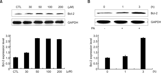
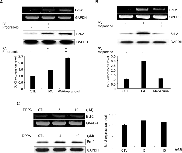
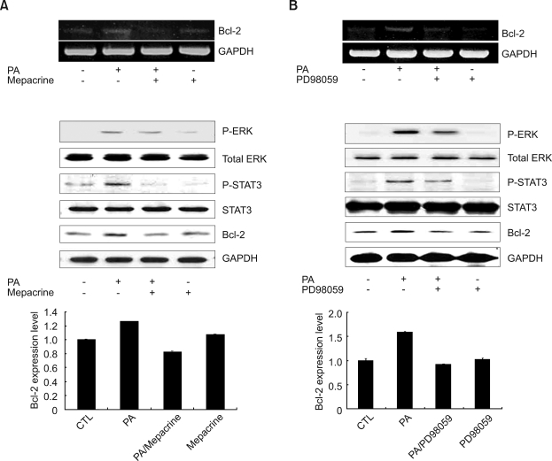
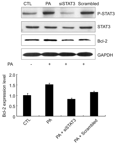
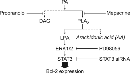
Phosphatidic acid (PA), the product of a PLD-mediated reaction, is a lipid second messenger that participates in various intracellular signaling events and is known to regulate a growing list of signaling proteins. We found that Bcl-2 was upregulated by PA treatment in HeLa cells. However, how PA upregulates Bcl-2 expression has not yet been studied. In this study, we tried to discover the mechanisms of Bcl-2 up-regulation by PA treatment in HeLa cells. Treatment with PA resulted in significantly increased expression of Bcl-2 in HeLa cells. Moreover, PA-induced Bcl-2 expression was blocked by mepacrine, an inhibitor of PLA2, but not by propranolol, an inhibitor of PA phospholyhydrolase (PAP). Treatment of 1,2-dipalmitoryl-sn-glycero-3- phosphate (DPPA) also increased Bcl-2 expression. These results indicate that Bcl-2 expression is mediated by lysophosphatidic acid (LPA), not by arachidonic acid (AA). Thereafter, we used MEK1/2 inhibitor, PD98059 to investigate the relationship between ERK1/2 MAPK and PA-induced Bcl-2 expression. PA-induced Bcl-2 expression was decreased when ERK1/2 was inhibited by PD98059. The transcription factor such as STAT3 which is controlled by ERK1/2 MAPK was increased along with Bcl-2 expression when the cells were treated with PA. Furthermore, STAT3 siRNA treatments inhibited PA-induced Bcl-2 expression, suggesting that STAT3 (Ser727) is involved in PA-induced Bcl-2 expression. Taken together, these findings indicate that PA acts as an important mediator for increasing Bcl-2 expression through STAT3 (Ser727) activation via the ERK1/2 MAPK pathway.


Figures

Figure 1
Figure 1
Effects of PA on the Bcl-2 expression in HeLa cells. PA increased Bcl-2 expression in both a dose- and time-dependent manner. (A) HeLa cells were cultured in DMEM containing 10% FBS; HeLa cells were further incubated without FBS for 18 h and treated with different concentrations of PA for 3 h. (B) Cells treated with 50 µM PA for the indicated time. The relative quantities of each protein band, normalized to control cells, were quantified using Quantity One software (Bio-Rad).
Figure 2
Figure 2
Effects of propranolol, mepacrine, and DPPA pretreatment on the expression of Bcl-2 in HeLa cells. (A) HeLa cells were pretreated with 50 µM propranolol for 30 min before being treated with 50 µM PA for 30 min for RT-PCR, and for 3 h for Western blotting. (B) HeLa cells were pretreated with 50 µM mepacrine for 30 min before 50 µM PA treatmemt for 30 min for RT-PCR, and for 3 h for Western blotting, respectively. (C) HeLa cells were cultured for 1 h with 5 and 10 µM DPPA after starvation for 18 h, respectively. The upper panel represents mRNA expression and lower panel represents protein. The cells were harvested, lysised and subjected to RT-PCR and Western blotting as described in the Methods. The relative quantities of each protein band, normalized to control cells, were quantified using Quantity One software (Bio-Rad).
Figure 3
Figure 3
Effects of a PLA2 inhibitor, mepacrine, and MEK inhibitor, PD98059, on PA-induced ERK1/2, STAT3 (Ser727) phosphorylation, and Bcl-2 expression in HeLa cells. (A) RNA was extracted from cells pretreated with 50 µM mepacrine for 30 min, followed by stimulation with 50 µM PA for 30 min and Bcl-2 mRNA was amplified by RT-PCR. HeLa cells were pretreated with 50 µM mepacrine for 30 min, followed by stimulation with 50 µM PA for 15 min. Cell lysates were analyzed by immunoblotting with anti-ERK1/2, anti-STAT3, and phospho-ERK1/2, phospho-STAT3 (Ser727) antibodies. In the case of Bcl-2 Western blotting, cells were pretreated with 50 µM mepacrine for 30 min, followed by stimulation with 50 µM PA for 3 h. Cell lysates were analyzed by immunoblotting with Bcl-2 antibody. (B) RNA was extracted from cells treated with 50 µM PD98059 for 1 h, followed by stimulation with 50 µM PA for 30 min and Bcl-2 mRNA was amplified by RT-PCR. HeLa cells were pretreated with 50 µM PD98059 for 1 h, followed by stimulation with 50 µM PA for 15 min. Cell lysates were analyzed by immunoblotting with anti-ERK1/2, STAT3, and phospho-ERK1/2, STAT3 (Ser727) antibodies. In the case of Bcl-2 Western blotting, cells were pretreated with 50 µM PD98059 for 1 h, followed by stimulation with 50 µM PA for 3 h. Cell lysates were analyzed by immunoblotting with Bcl-2 antibody. The relative quantities of each protein band, normalized to control cells, were quantified using Quantity One software (Bio-Rad).
Figure 4
Figure 4
Effects of STAT3 siRNA on the expression of Bcl-2 in HeLa cells. HeLa cells were transiently transfected with 100 nM STAT3 siRNA or scramble siRNA for 72 h and then stimulated with 50 µM PA for 15 min (for p-STAT3/STAT3 blots) or 3 h (for Bcl-2 blots). Expression level of p-STAT3 (Ser727), STAT3, and Bcl-2 were determined by Western blot analysis. The relative quantities of each protein band, normalized to control cells, were quantified using Quantity One software (Bio-Rad).
Figure 5
Figure 5
A proposed model for the signaling pathway of PA-induced Bcl-2 expression. We diagramed a mechanism of up-regulation of Bcl-2 expression induced by PA. PA can be converted to LPA by PLA2, but does not pass through the DAG pathway by PAP. Subsequently, LPA acts as an important signal molecule to upregulate Bcl-2 expression. PA leads to activation of downstream kinases, ERK1/2, which are responsible for the phosphorylation of STAT3 (Ser727).

References

    1. Adams JM, Cory S. Life-or-death decisions by the Bcl-2 protein family. Trends Biochem Sci. 2001;26:61–66. - PubMed
    1. Alas S, Bonavida B. Rituximab inactivates signal transducer and activation of transcription 3 (STAT3) activity in B-non-Hodgkin's lymphoma through inhibition of the interleukin 10 autocrine/paracrine loop and results in down-regulation of Bcl-2 and sensitization to cytotoxic drugs. Cancer Res. 2001;61:5137–5144. - PubMed
    1. Boucher MJ, Morisset J, Vachon PH, Reed JC, Laine J, Rivard N. MEK/ERK signaling pathway regulates the expression of Bcl-2, Bcl-X (L), and Mcl-1 and promotes survival of human pancreatic cancer cells. J Cell Biochem. 2000;79:355–369. - PubMed
    1. Bromberg JF, Wrzeszczynska MH, Devgan G, Zhao Y, Pestell RG, Albanese C, Darnell JE., Jr Stat3 as an oncogene. Cell. 1999;98:295–303. - PubMed
    1. Cho JH, Hong SK, Kim EY, Park SY, Park CH, Kim JM, Kwon OJ, Kwon SJ, Lee KS, Han JS. Overexpression of phospholipase D suppresses taxotere-induced cell death in stomach cancer cells. Biochim Biophys Acta. 2008;1783:912–923. - PubMed

Publication types

MeSH terms

LinkOut - more resources