

Characterization of brain development in the ferret via MRI
- PMID: 19287340
- PMCID: PMC3384539
- DOI: 10.1203/PDR.0b013e3181a291d9
Characterization of brain development in the ferret via MRI
Abstract
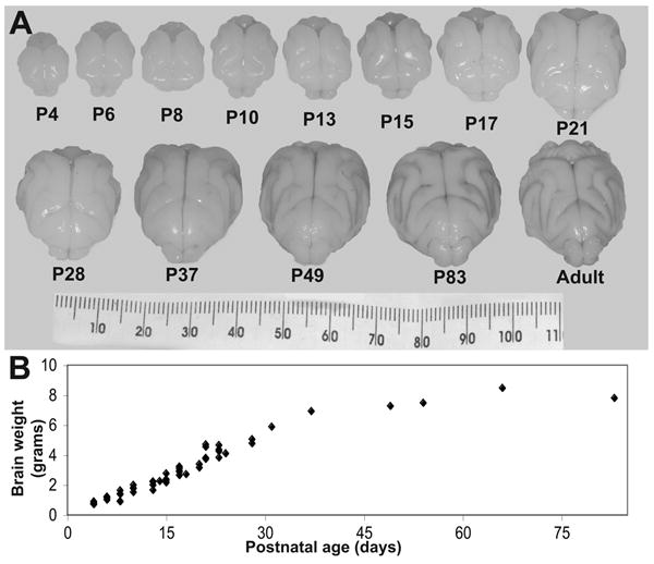
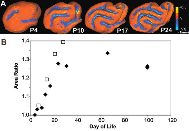
Animal models with complex cortical development are useful for improving our understanding of the wide spectrum of neurodevelopmental challenges facing human preterm infants. MRI techniques can define both cerebral injury and alterations in cerebral development with translation between animal models and the human infant. We hypothesized that the immature ferret would display a similar sequence of brain development [both gray (GM) and white matter (WM)] to that of the preterm human infant. We describe postnatal ferret neurodevelopment with conventional and diffusion MRI. The ferret is born lissencephalic with a thin cortical plate and relatively large ventricles. Cortical folding and WM maturation take place during the first month of life. From the mid-second through the third week of postnatal life, the ferret brain undergoes a similar, though less complex, pattern of maturational changes to those observed in the human brain during the second half of gestation. GM anisotropy decreases rapidly in the first 3 wks of life, followed by an upward surge of surface folding and WM anisotropy over the next 2 wks.
Figures


References
-
- Moster D, Lie RT, Markestad T. Long-term medical and social consequences of preterm birth. N Engl J Med. 2008;359:262–273. - PubMed
-
- Back SA, Riddle A, Hohimer AR. Role of instrumented fetal sheep preparations in defining the pathogenesis of human periventricular white-matter injury. J Child Neurol. 2006;21:582–589. - PubMed
-
- Derrick M, Drobyshevsky A, Ji X, Tan S. A model of cerebral palsy from fetal hypoxia-ischemia. Stroke. 2007;38:731–735. - PubMed
-
- Dieni S, Inder T, Yoder B, Briscoe T, Camm E, Egan G, Denton D, Rees S. The pattern of cerebral injury in a primate model of preterm birth and neonatal intensive care. J Neuropathol Exp Neurol. 2004;63:1297–1309. - PubMed
-
- Duncan JR, Cock ML, Scheerlinck JP, Westcott KT, McLean C, Harding R, Rees SM. White matter injury after repeated endotoxin exposure in the preterm ovine fetus. Pediatr Res. 2002;52:941–949. - PubMed
Publication types
MeSH terms
Grants and funding
LinkOut - more resources
Full Text Sources
