. 2009 Apr;11(4):451-9.
doi: 10.1038/ncb1852. Epub 2009 Mar 15.

p53-cofactor JMY is a multifunctional actin nucleation factor

p53-cofactor JMY is a multifunctional actin nucleation factor

J Bradley Zuchero et al. Nat Cell Biol. 2009 Apr.

Abstract

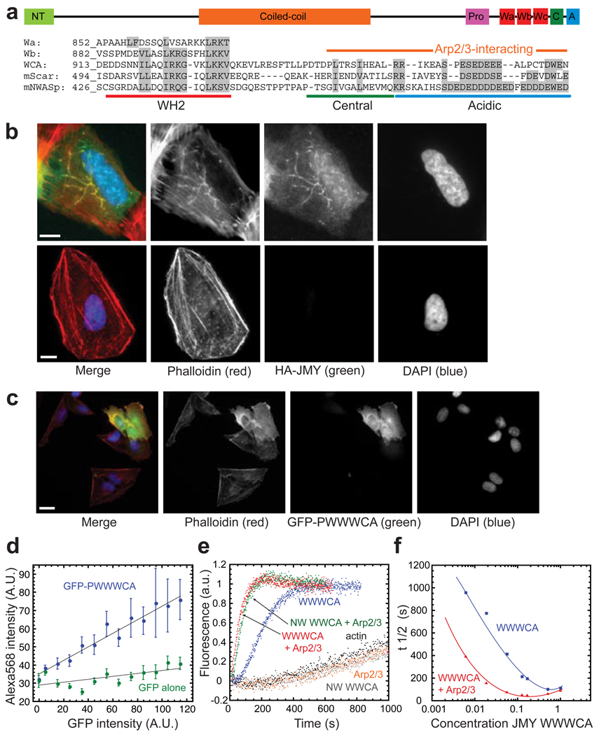
Many cellular structures are assembled from networks of actin filaments, and the architecture of these networks depends on the mechanism by which the filaments are formed. Several classes of proteins are known to assemble new filaments, including the Arp2/3 complex, which creates branched filament networks, and Spire, which creates unbranched filaments. We find that JMY, a vertebrate protein first identified as a transcriptional co-activator of p53, combines these two nucleating activities by both activating Arp2/3 and assembling filaments directly using a Spire-like mechanism. Increased levels of JMY expression enhance motility, whereas loss of JMY slows cell migration. When slowly migrating HL-60 cells are differentiated into highly motile neutrophil-like cells, JMY moves from the nucleus to the cytoplasm and is concentrated at the leading edge. Thus, JMY represents a new class of multifunctional actin assembly factor whose activity is regulated, at least in part, by sequestration in the nucleus.


Figures

Figure 1
Figure 1. JMY nucleates actin filaments and activates the Arp2/3 complex
(a) Domain structure of JMY. The C-terminus of JMY is homologous to activators of Arp2/3. A poly-proline (P) domain is followed by three tandem actin monomer-binding WH2 domains (Wa through Wc), an actin and Arp2/3-binding central domain (C), and an Arp2/3-binding acidic domain (A). Alignment shows individual WH2 domains of JMY, and compares the sequences of the WCA regions of JMY, Scar, and N-WASp. JMY WCA is 28% identical to N-WASp WCA (ClustalW), and residues putatively involved in binding actin and Arp2/3, are 100% conserved between all available JMY sequences. (b) Expression of HA-JMY (top panels; visualized by indirect immunofluorescence of HA, green) in U2OS cells induces the formation of filamentous actin structures (visualized with Alexa Fluor 568 phalloidin, red). These elongated actin structures colocalize with JMY and are not seen in untransfected cells (bottom panels). Nuclei were visualized with DAPI (blue). Scale bar, 10 µm. (c) Expression of GFP-PWWWCA (green) in U2OS cells increases cellular F-actin (Alexa Fluor 568 phalloidin, red). (d) Quantification of the increase in F-actin induced by GFP-PWWWCA expression. Phalloidin intensity was plotted as a function of GFP-PWWWCA intensity and shows a linear increase in F-actin content with increased expression of GFP-PWWWCA (n=457). In contrast, expressing GFP alone has only a minor effect on the red intensity detected, likely due to a small amount of bleed-through (n=458). (e) Pyrene-actin polymerization assays show that JMY WWWCA both activates Arp2/3 (R) and nucleates actin in the absence of Arp2/3 (red and blue traces). N-WASp (NW) WWCA activates Arp2/3 (green), but does not nucleate actin on its own (grey). (f) Intrinsic nucleation and activation of Arp2/3 by WWWCA are dose-dependent. Pyrene-actin polymerization assays were conducted in the absence (blue) or presence (red) of Arp2/3, with increasing concentrations of JMY WWWCA. Time to half-maximal polymerization was plotted as a function of WWWCA concentration. Pyrene-actin polymerization assays were in 1x KMEI and contained 2 µM actin, 167 nM JMY or N-WASp, and 2.5 nM Arp2/3, where noted.
Figure 2
Figure 2. Mechanistic dissection of JMY
(a) JMY nucleates unbranched filaments, and increases the number of filaments over actin alone. Filaments made in the presence (left) or absence (right) of 167 nM JMY WWWCA were fixed with Alexa Fluor 488 phalloidin and Latrunculin B at 6 minutes (t9/10 of JMY WWWCA reaction) prior to dilution and spotting on poly-L-lysine coverslips,. Scale bars, 5 µm. (b) Quantification of filaments per field in images from a demonstrates that JMY nucleates new filaments (JMY, 42.7 ± 4.2 filaments per micron, n=35 fields; actin alone, 3.2 ± 0.5 filaments per micron, n=30 fields). (c) Filaments prepared as in a, in the presence of JMY plus Arp2/3 (left), Scar plus Arp2/3 (centre), or actin alone (right). The concentration of filaments was kept constant by arresting reactions at their individual t9/10s (see Methods). Filaments nucleated in the presence of JMY and Arp2/3 are branched, consistent with JMY activating Arp2/3, as are filaments made in the presence of Scar and Arp2/3. The shorter, more abundant filaments seen here are due to Arp2/3 nucleating actin more rapidly than intrinsic nucleation by JMY. (d) Quantification of filament length at t9/10. The rate of nucleation is inversely proportional to the length of filament (rate: JMY+Arp2/3 > JMY > Scar+Arp2/3 ≫ actin alone). (e) Quantification of filament branching in each condition. n>300 filaments per condition. (f) Tandem WH2 domains from JMY are sufficient for actin nucleation. Actin polymerization is as fast with WWW as it is with WWWCA. WWW lacks the Arp2/3 binding CA domain, so adding Arp2/3 to WWW does not accelerate polymerization over WWW alone. N-WASp WW does not nucleate actin. (g) JMY WCA is sufficient to activate Arp2/3, but does not nucleate actin. The rate of actin polymerization in the presence of JMY WCA and Arp2/3 is similar to reactions containing Scar WCA and Arp2/3. In the absence of Arp2/3, JMY WCA has no effect on actin polymerization. Experimental conditions as in Fig. 1.
Figure 3
Figure 3. JMY nucleates actin by the same mechanism as Spire
(a) The region between JMY Wb and Wc is homologous to the short actin nucleation motif from Spire (monomer-binding linker, MBL). The figure shows an alignment between Spire-MBL and the same region of JMY (mJMY AA 903–917) with homologous residues coloured grey. The MBL sequence is not homologous to the analogous position in N-WASp, WHAMM, or Cordon Bleu. The position of the glycine-serine repeats in b is underlined, and the sequences of N-WASp gain of function mutations (NW WJW and WSW) in c are shown. (b) JMY-MBL is important for actin nucleation. Replacing the MBL in JMY WbWc and Spire CD (the two C-terminal WH2 domains in Spire) with a flexible linker of glycine-serine repeats (gs5) causes a nucleation defect in both JMY and Spire. The analogous N-WASp mutant does not promote nucleation, but inhibits spontaneous polymerization. (c) Gain of function. Replacing the linker region between the WH2 domains of N-WASp WW with JMY- or Spire-MBL (NW WJW or WSW) converts N-WASp into a weak actin nucleator. This shows that JMY- and Spire-MBL are sufficient for nucleation. Mutated amino acids of WJW and WSW are shown in a. Reactions in b and c contained 4 µM actin. Experimental conditions as in Fig. 1. (d) t/12s of reactions from b–c. Reactions were repeated > 3 times each. Error bars, s.e.m. (e) Actin nucleation and activation of Arp2/3 are distinct activities of JMY that overlap spatially. Tandem JMY WH2 domains and the MBL (star in figure) nucleate actin, similar to Spire, and JMY WCA activates Arp2/3, similar to N-WASp and Scar.
Figure 4
Figure 4. JMY localizes to the leading edge of motile cells
(a–c) Redistribution of JMY from the nucleus to the leading edge in HL-60 cells. (a) JMY is primarily nuclear in undifferentiated HL-60 cells. (b) Following differentiation into motile cells by culturing in 1.3% DMSO for 5–7 days, JMY colocalizes with filamentous actin in the cytoplasm. (c) Differentiated HL-60 cells were polarized by exposure to 100 nM fMLP, a chemoattractant. JMY is distributed throughout the cytoplasm, where it colocalizes strongly with filamentous actin at the leading edge. Cells were fixed and stained with Alexa Fluor 568 phalloidin (red), anti-JMY (green), and DAPI (blue). Scale bars, 10 µm. (d) Human primary neutrophils were obtained by finger pinprick, stimulated with 20 nM fMLP, and fixed and stained as above. JMY (green) colocalizes with filamentous actin (red) at the leading edge. Scale bar, 5 µm. (e) Western blots of JMY and binding partner p300 in undifferentiated (U) and differentiated (D) HL-60 cells. p300 is expressed in undifferentiated, but not differentiated, HL-60 cells.
Figure 5
Figure 5. JMY contributes to cell motility
(a–b) Expressing GFP-JMY in U2OS cells significantly increases their motility in wound healing assays. Stable lines of GFP-JMY and GFP-JMYΔCA were wounded by scraping with micropipette tips. Images were acquired at 0, 2, 4, 6, and 12 hours after wounding. (b) Migration rate from 0–6 hours was averaged from a minimum of 4 replicates on each of 3 days. GFP-JMY expression induces cells to migrate 16.6% faster than wild-type cells (n=3, p<0.003). Cells expressing a truncation of JMY lacking the Arp2/3-interacting CA domain migrate at the same rate as wild-type cells. Scale bars, 100 µm. (c–e) Wound healing assays in U2OS cells indicate that knocking-down JMY by RNAi impairs cell migration. Cells were transfected with JMY or control non-targeting 2 (Dharmacon) siRNA (C) and wounded (red dashed line) as in a. Images were taken at the same position 0, 3, 6, and 24 h after wounding. (d) Wound size at each time point for all conditions. (n=4). (e) Western blots show RNAi efficiency in U2OS cells. (f) Knocking down JMY expression decreases cellular levels of F-actin. HEK 293 cells were transfected with a vector encoding both GFP and a JMY-specific shRNA and fixed and stained with Alexa Fluor 568-phalloidin. Average phalloidin intensity in GFP-negative (non-RNAied) and GFP-positive (RNAied) cells is plotted (n=396, p<0.03, see Methods). Error bars, s.e.m. (g) JMY localizes to the leading edge of U2OS cells. Cells were grown as above, and fixed and stained for JMY 15 minutes after wounding. JMY is primarily nuclear, but it is also enriched at the leading edge (indirect immunofluorescence, green) where it colocalizes with a subset of actin filaments (Alexa Fluor 568-phalloidin, red). Inset: leading edge, contrast enhanced to show actin filaments and JMY. Scale bar, 10 µm. (h) Models of in vivo role of JMY. Top, Model 1: JMY nucleates filaments that then serve as substrates for dendritic nucleation by Arp2/3. Bottom, Model 2: JMY evolved to nucleate actin in different cellular contexts. In the nucleus it nucleates unbranched filaments, and in the cytoplasm it both nucleates filaments and activates Arp2/3.

Comment in

  • Double JMY: making actin fast.
    Roadcap DW, Bear JE. Roadcap DW, et al. Nat Cell Biol. 2009 Apr;11(4):375-6. doi: 10.1038/ncb0409-375. Nat Cell Biol. 2009. PMID: 19337319 Free PMC article.

References

    1. Welch M, Mullins R. Cellular control of actin nucleation. Annu. Rev. Cell Dev. Biol. 2002;18:247–288. - PubMed
    1. Quinlan ME, Heuser JE, Kerkhoff E, Mullins RD. Drosophila Spire is an actin nucleation factor. Nature. 2005;433:382–388. - PubMed
    1. Marchand JB, Kaiser DA, Pollard TD, Higgs HN. Interaction of WASP/Scar proteins with actin and vertebrate Arp2/3 complex. Nat Cell Biol. 2001;3:76–82. - PubMed
    1. Shikama N, et al. A novel cofactor for p300 that regulates the p53 response. Mol. Cell. 1999;4:365–376. - PubMed
    1. Coutts A, Boulahbel H, Graham A, La Thangue N. Mdm2 targets the p53 transcription cofactor JMY for degradation. EMBO Rep. 2006;8:84–90. - PMC - PubMed

Publication types