. 2009 Apr;12(4):409-17.
doi: 10.1038/nn.2287. Epub 2009 Mar 15.

Proteoglycan interactions with Sonic Hedgehog specify mitogenic responses

Affiliations

Proteoglycan interactions with Sonic Hedgehog specify mitogenic responses

Jennifer A Chan et al. Nat Neurosci. 2009 Apr.

Abstract

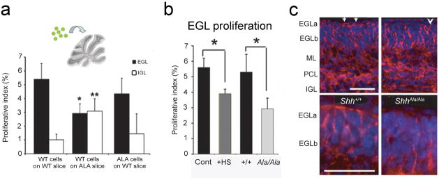
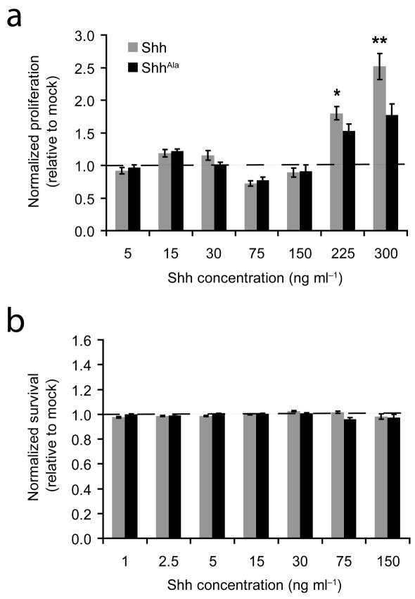
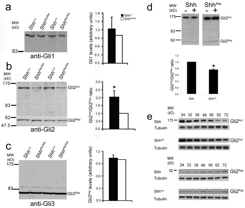
Sonic Hedgehog (Shh) has dual roles in vertebrate development, promoting progenitor cell proliferation and inducing tissue patterning. We found that the mitogenic and patterning functions of Shh can be uncoupled from one another. Using a genetic approach to selectively inhibit Shh-proteoglycan interactions in a mouse model, we found that binding of Shh to proteoglycans was required for proliferation of neural stem/precursor cells, but not for tissue patterning. Shh-proteoglycan interactions regulated both spatial and temporal features of Shh signaling. Proteoglycans localized Shh to specialized mitogenic niches and also acted at the single-cell level to regulate the duration of Shh signaling, thereby promoting a gene expression program that is important for cell division. Because activation of the Shh pathway is a feature of diverse human cancers, selective stimulation of proliferation by Shh-proteoglycan interactions may also figure prominently in neoplastic growth.


Figures

Figure 1
Figure 1. ShhAla specifically alters proteoglycan binding
(a) ShhAla shows reduced binding to heparin-coated plates. Shh-AP (circles), ShhAla-AP (squares), or AP (triangles) were incubated with heparin-coated plates in the presence of increasing concentrations of soluble heparin. (b) Shh binds GPI-linked proteoglycans in cerebellar sections; ShhAla does not. P6 sections treated with vehicle control (−), Heparinases or PI-PLC, incubated with Shh-AP, ShhAla-AP or vehicle controls, and processed for binding of AP-tagged ligand. Scale bar, 100 μm. (c) ShhAla is processed to mature isoform. Lysates of HEK293 transfected with Shh or ShhAla were analyzed by immunoblot with anti-Shh. Immature 45kD (arrowhead) and mature 20kD isoform (arrow) are seen. (d) ShhAla is palmitoylated. HEK293 cells expressing Shh, ShhAla or ShhC24S were labeled with 3H-palmitate, analyzed for palmitoylation, and probed with anti-Shh. Shh and ShhAla are palmitoylated. ShhC24S is not.
Figure 2
Figure 2. ShhAla/Ala and ShhAla/− mice exhibit defects in growth, with normal patterning
(a) Skeletal morphology, body and brain patterning are normal in ShhAla/Ala mice, but size of adult ShhAla/Ala mice is 11% less than wild type, and sizes of olfactory bulbs and cerebella are 30 and 31% reduced (arrows) respectively. Sagittal view of cerebellum shows normal patterning with reduced size in ShhAla/Ala. Scale bar, 1 mm. ShhAla/Ala animals also display well-spaced eyes. (b) Spinal cord patterning is normal in ShhAla/Ala mice. In situ hybridization for Shh (expressed in notochord and floor plate), Nkx2.2 (motor neuron precursors), Nkx6.1 (ventral spinal cord), Isl1 (dorsal root ganglia and motor neurons), and Dbx1 (V0 interneurons) in E10.5 littermates. Scale bar, 100 μm. (c) Growth defects are seen in ShhAla/−, without defects in patterning. Olfactory bulb and cerebellum are reduced in size (red arrows). Scale bar, 1 mm. Eyes of ShhAla/− mice are well spaced.
Figure 3
Figure 3. Reduced proliferation of ShhAla/Ala cerebellar granule precursors is seen in the EGL of developing mice
(a) p-Histone H3 (p-H3)-positive or BrdU (BrdU)-positive (both in red) cells are fewer in the EGL of P3 ShhAla/Ala mice as compared to Shh+/+ littermates. DAPI is in grey or blue. Scale bar, 100 μm (left), 50 μm (right). (b) p-Histone H3 mitotic indices are reduced in ShhAla/Ala mice (white bars) at multiple postnatal ages (Shh+/+, black bars) (*p<0.001). Reduced proliferation in ShhAla/Ala mice is also seen in one wild type and mutant littermate pair at P9 (1.37% versus 1.07% for Shh+/+ and ShhAla/Ala, respectively). Error bars are +/− s.e.m. (c) BrdU proliferation indices are less in ShhAla/Ala mice (white bars) as compared to wild type (black bars) at multiple postnatal ages (*p<0.001). While no difference is seen at intermediate time points (P6 and P9), this, in part, reflects the cerebellar size difference between ShhAla/Ala and Shh+/+ mice. There is still a significant difference in the number of proliferating cells/EGL length at P6 (3.6% in Shh+/+ versus 2.7% in ShhAla/Ala). Error bars are +/− s.e.m. (d) p-Histone H3 mitotic indices in P3 wild type (black bars) and ShhAla/Ala mice (white bars) in the anterior, middle and posterior cerebellum are not statistically different from their respective totals in (b), demonstrating that lobes throughout the cerebellum are affected in the mutant. Error bars are +/− s.e.m.
Figure 4
Figure 4. Embryonic and adult neural stem/precursor proliferation is reduced in ShhAla/Ala mice
(a) More proliferating neural stem/precursors are seen in the spinal cord of Shh+/+ mice (left) than in their ShhAla/Ala (right) E10.5 littermates. p-Histone H3 is in red and DAPI in blue. Scale bar, 100 μm. (b) A reduction in the total number of p-Histone H3 positive (p-H3+) cells in the spinal cord of ShhAla/Ala embryos is seen compared to the number in ShhCtl/Ctl littermates (*p<0.05). A significant reduction in the total p-H3+ cells per unit spinal cord area is also seen in ShhAla/Ala E10.5 embryos (data not shown). Error bars are +/− s.e.m. (c) Fewer p-H3+ cells are present in the SVZ of adult ShhAla/Ala mice (*p<0.05). Error bars are +/− s.e.m. (d) Fewer BrdU positive (BrdU+) cells are seen in the hippocampal SGL of adult mutant mice (*p≤0.05). Error bars are +/− s.e.m.
Figure 5
Figure 5. ShhAla cannot specify a mitogenic niche
(a) Granule cell precursors (GCPs) from GFP+ mice were cultured on P6 Shh+/+ or ShhAla/Ala cerebellar slices, or DiO-labeled ShhAla/Ala GCPs were cultured on wild type slices. Proliferation of introduced GCPs was analyzed by BrdU incorporation. The proliferative index is the percent (%) of GFP- or DiO-positive cells in each layer that are BrdU+ (*p<0.05 versus EGL of WT slice; **p<0.05 vs IGL of WT slice; no significant difference between EGL and IGL proliferation on ShhAla/Ala slices is seen). Error bars are +/− s.e.m. (b) EGL mitogenic niche in ShhAla/Ala slices (light gray bar) is phenocopied by added exogenous heparan sulfates (HS) (dark gray bar) (*p<0.05). Error bars are +/− s.e.m. (c) Shh immunostaining of wild type and ShhAla/Ala P6 cerebella shows a reduction in Shh staining in the EGLa of mutant mice (arrowhead) compared to wild type cerebella (arrows) (52%±11% of wild type (p=0.01)). EGLa: external granule cell layer, outer proliferative zone, EGLb: external granule cell layer, inner post-mitotic zone; ML: molecular layer; PCL: Purkinje cell layer; IGL: internal granule cell layer. Scale bar, 50 μm
Figure 6
Figure 6. Shh-proteoglycan interactions promote proliferation in dissociated cell cultures of GCPs, but are not needed for survival
(a) The proliferative response to Shh (gray) (as assessed by BrdU incorporation) is greater than that of GCPs to ShhAla (black) (*p≤0.05, **p<0.01). Error bars are +/− s.e.m. (b) No consistent effects on survival (as assessed by activated caspase 3 immunostaining) are seen in GCPs stimulated with Shh (gray) or ShhAla (black). Error bars are +/− s.e.m.
Figure 7
Figure 7. Shh-proteoglycan interactions modulate transcriptional activity through the regulation of Gli2 isoforms and signaling kinetics
(a) Gli1 protein levels in P3 ShhAla/Ala and Shh+/+ cerebella are equivalent. At left, anti-Gli1 western blot of ShhAla/Ala and Shh+/+ cerebellar lysates. At right, quantification of blotting results, ShhAla/Ala (white bar), Shh+/+ (black bar). Error bars are +/− s.e.m. (b) The ratio of Gli2 activator (Gli2Act) to repressor (Gli2Rep) is reduced in P3 ShhAla/Ala cerebella compared to Shh+/+. At left, anti-Gli2 western blot. At right, quantification of results, ShhAla/Ala (white bar), Shh+/+ (black bar) (*p<0.01). Error bars are +/− s.e.m. (c) Gli3 repressor protein levels are unchanged in P3 ShhAla/Ala cerebella, as compared to Shh+/+. At left, anti-Gli3 western blot. At right, quantification of results, ShhAla/Ala (white bar), Shh+/+ (black bar). Error bars are +/− s.e.m. (d) The ratio of Gli2 activator (Gli2Act) to repressor (Gli2Rep) is greater in cells stimulated with Shh compared to ShhAla. At top, anti-Gli2 western blot. At bottom, quantification of results, Shh (left bar), ShhAla (right bar) (*p<0.01). Error bars are +/− s.e.m. (e) Kinetics of signaling differ for GCPs stimulated with ShhAla as compared to GCPs stimulated with Shh. At top, anti-Gli2Act western blot of Shh- or ShhAla-stimulated GCPs (or blot against tubulin as a loading control). Numbers above blots are hours in culture. At bottom, anti-Gli2Rep western blot of Shh- or ShhAla-stimulated GCPs (or blot against tubulin as a loading control).
Figure 8
Figure 8. Proteoglycan interactions modulate Shh perdurance and differentially affect Shh-dependent gene expression
(a) The gene pattern induced by ShhAla stimulation of wild type P6 GCPs is different than that of GCPs stimulated by equivalent amounts of Shh (*p<0.05). Error bars are +/− s.e.m. (b) GCP proteoglycans modulate Shh ligand perdurance. At left, (top) anti-Shh western blot of Shh- or ShhAla-stimulated C3H10T1/2s (or blot against actin as loading control). At right, (top) anti-Shh western blot of Shh- or ShhAla-stimulated GCPs (or blot against tubulin as loading control). Numbers above blots are hours in culture. At bottom, quantification of results from one representative experiment. Shh (solid), ShhAla (dashed). (c) Expression of Shh target genes in P1-2 ShhAla/Ala cerebella (P1-3 ShhAla/Ala cerebella for gli2) relative to Shh+/+ littermates demonstrates that Shh-proteoglycan interactions differentially affect gene subsets (*p<0.01, **p<0.05). Error bars are +/− s.e.m.

Comment in

References

    1. Capdevila J, Izpisua Belmonte JC. Patterning mechanisms controlling vertebrate limb development. Annu Rev Cell Dev Biol. 2001;17:87–132. - PubMed
    1. Hooper JE, Scott MP. Communicating with Hedgehogs. Nature reviews. 2005;6:306–317. - PubMed
    1. Machold R, et al. Sonic hedgehog is required for progenitor cell maintenance in telencephalic stem cell niches. Neuron. 2003;39:937–950. - PubMed
    1. Palma V, et al. Sonic hedgehog controls stem cell behavior in the postnatal and adult brain. Development. 2005;132:335–344. - PMC - PubMed
    1. Bellaiche Y, The I, Perrimon N. Tout-velu is a Drosophila homologue of the putative tumour suppressor EXT-1 and is needed for Hh diffusion. Nature. 1998;394:85–88. - PubMed

Publication types

MeSH terms