. 2009 Apr;16(4):397-404.
doi: 10.1038/nsmb.1572. Epub 2009 Mar 15.

The pathway of hepatitis C virus mRNA recruitment to the human ribosome

Affiliations

The pathway of hepatitis C virus mRNA recruitment to the human ribosome

Christopher S Fraser et al. Nat Struct Mol Biol. 2009 Apr.

Abstract

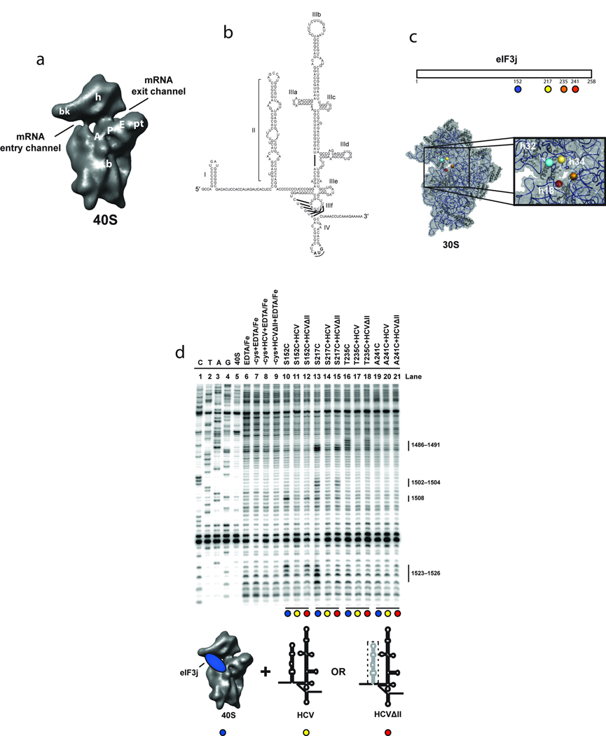
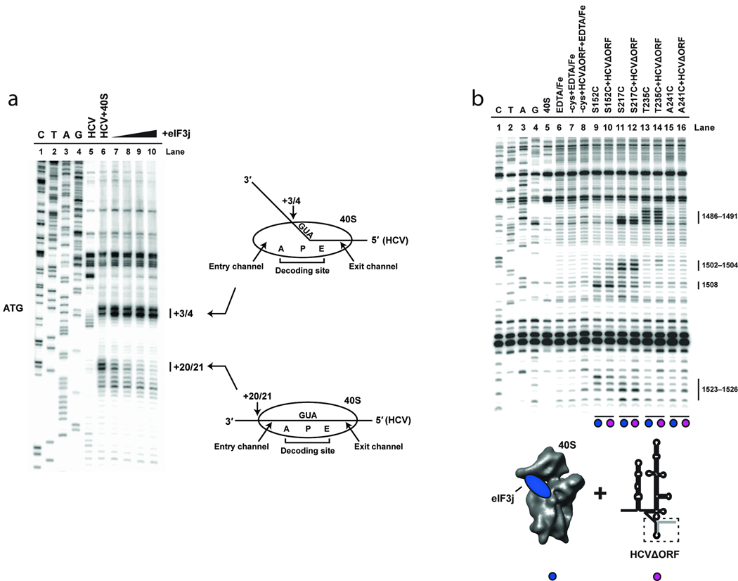
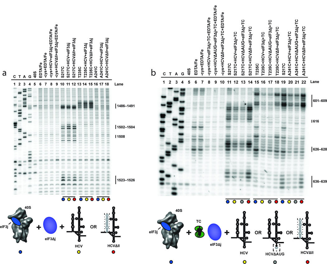
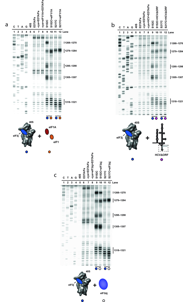
Eukaryotic protein synthesis begins with mRNA positioning in the ribosomal decoding channel in a process typically controlled by translation-initiation factors. Some viruses use an internal ribosome entry site (IRES) in their mRNA to harness ribosomes independently of initiation factors. We show here that a ribosome conformational change that is induced upon hepatitis C viral IRES binding is necessary but not sufficient for correct mRNA positioning. Using directed hydroxyl radical probing to monitor the assembly of IRES-containing translation-initiation complexes, we have defined a crucial step in which mRNA is stabilized upon initiator tRNA binding. Unexpectedly, however, this stabilization occurs independently of the AUG codon, underscoring the importance of initiation factor-mediated interactions that influence the configuration of the decoding channel. These results reveal how an IRES RNA supplants some, but not all, of the functions normally carried out by protein factors during initiation of protein synthesis.


Figures

Figure 1
Figure 1
Directed hydroxyl radical probing of 18S rRNA from BABE-Fe-eIF3j-40S-HCV complexes. (a) The 40S subunit structure based on a cryo-EM reconstruction viewed from the subunit interface with landmarks indicated: A, A-site; P, P-site; E, E-site; bk, beak; b, body; pt, platform; and h, head. (b) The 5´ UTR of the HCV mRNA consists of four domains (I–IV); the IRES domains (II–IV) with sub-domains (a–f) of domain III are indicated. (c) Representation of eIF3j indicating the positions of cysteine mutations used for BABE-Fe attachment (upper panel). Modeled positions of eIF3j amino acids in the T. thermophilus 30S crystal structure, adapted from a previous publication (lower panel). The boxed area provides a detailed view of the mRNA entry channel and A-site with helices 18, 32 and 34 indicated. Nucleotides cleaved in these helices for each experiment are shown in Supplementary Fig. 1a. (d) Primer extension analysis of 18S rRNA cleaved by BABE-Fe-modified eIF3j. Sequencing lanes are indicated (C, T, A, and G). Other control lanes include 40S subunits in the absence or presence of EDTA/Fe, mock-derivatized eIF3j (−cys+EDTA-Fe) in the absence (lane 7) or presence of wild type (HCV; lane 8), or domains III-IV (HCVΔII ; lane 9) of the HCV IRES RNA. Other lanes include eIF3j derivatized with BABE-Fe at the positions indicated either in the absence (lanes 10, 13, 16, and 19), or presence of HCV IRES (lanes 11, 14, 17, and 20), or HCVΔII IRES (lanes 12, 15, 18, and 21). 18S rRNA nucleotide positions of cleavage sites are indicated. Colored circles indicate components added in each reaction as depicted in the cartoon. The deletion of domain II (HCVΔII) is represented by a dotted line.
Figure 2
Figure 2
Toeprinting analysis of the 40S-HCV-eIF3j complexes. (a) Lanes C, T, A, and G depict sequencing lanes corresponding to HCV mRNA, with the AUG codon indicated. Toeprinting reactions of HCV mRNA in the absence (lane 5) or presence (lane 6) of 40S subunits is shown. Additional reactions including 40S subunits in the presence of 5 µM (lane7), 10 µM (lane 8), 20 µM (lane 9), or 40 µM (lane 10) eIF3j are indicated. The positions of toeprints that correspond to 40S-HCV complexes are indicated (+3/4 and +20/21). Numbering is from the A (+1) of the AUG codon. Cartoons depicting the 40S-HCV complexes formed are also indicated. (b) Primer extension analysis of 18S rRNA cleaved by BABE-Fe-modified eIF3j in the absence or presence of HCV IRES RNA truncated after the AUG codon (HCVΔORF). Sequencing and control lanes are as indicated as described in Fig. 1d). The lanes corresponding to eIF3j in the absence (lanes 9, 11, 13, and 15) or presence (lanes 10, 12, 14, and 16) of HCVΔORF are indicated. As described in Fig. 1d, the colored circles correspond to the components added in each reaction, as indicated by the cartoons.
Figure 3
Figure 3
Effects of eIF3 and eIF2-Met-tRNAi on directed hydroxyl radical probing of 18S rRNA with BABE-Fe-eIF3j. (a) Lanes include 40S subunits in the absence or presence of EDTA/Fe and mock-derivatized eIF3j (−cys+EDTA-Fe) in the absence or presence of HCV constructs and eIF3 complex without endogenous eIF3j (eIF3Δj). Lanes corresponding to eIF3j derivatized with BABE-Fe at the positions indicated in the absence (lanes 10, 13, and 16), or presence of eIF3Δj and wild type HCV IRES (HCV; lanes 11, 14, and 17), or domain III of the HCV IRES (HCVΔII; lanes 12, 15, and 18) are indicated. (b) Lanes include 40S subunits in the absence or presence of EDTA/Fe and mock-derivatized eIF3j (−cys+EDTA-Fe) in the absence or presence of HCV constructs and other initiation factors as indicated. Lanes corresponding to BABE-Fe-modified eIF3j at the positions indicated in the absence (lanes 11, 15, and 19) or presence of eIF3Δj, eIF2-Met-tRNAi (Ternary complex; TC) and HCV (lanes 12, 16, and 20), HCV\AUG (lanes 13, 17 and 21), or HCVΔII (lanes 14, 18, and 22) are indicated. For each gel, sequencing lanes (C, T, A, and G) and cleavage nucleotide positions in the 18S rRNA are indicated. Colored circles correspond to the components added, as depicted in the cartoons. Relevant mutations in each HCV IRES construct are represented by a dotted line.
Figure 4
Figure 4
Effects of eIF1, eIF1A, HCV and eIF3 and on directed hydroxyl radical probing of 18S rRNA from BABE-Fe-eIF3j. (a) Primer extension analysis of 18S rRNA cleaved by BABE-Fe-modified eIF3j in the absence (lanes 9 and 11) or presence of eIF1 and eIF1A (lanes 10 and 12). (b) Analysis of 18S rRNA cleaved by BABE-Fe-modified eIF3j in the absence (lanes 9 and 11) or presence of HCVΔORF (lanes 10 and 12). (c) Analysis of 18S rRNA cleavage by BABE-Fe-modified eIF3j in the absence (lanes 9 and 11) or presence of eIF3Δj (lanes 10 and 12). In each gel the sequencing lanes are indicated (C, T, A, and G). Other lanes include 40S subunits in the absence or presence of EDTA/Fe and mock-derivatized eIF3j (−cys+EDTA-Fe) in the absence or presence of HCV and other initiation factors as indicated. Cleavage nucleotide positions in the 18S rRNA are indicated and colored circles correspond to the components added in each reaction, as depicted in the cartoons.
Figure 5
Figure 5
The effect of eIF2-Met-tRNAi on directed hydroxyl radical probing of 18S rRNA with BABE-Fe-eIF3j. Primer extension analysis of 18S rRNA cleaved by BABE-Fe-modified eIF3j. The sequencing lanes are indicated (C, T, A, and G). Other lanes include 40S subunits in the absence or presence of EDTA/Fe and mock-derivatized eIF3j (-cys+EDTA-Fe) in the absence (lane7) or presence of eIF2-Met-tRNAi (TC; lane 8). Lanes corresponding to eIF3j derivatized with BABE-Fe at the positions indicated in the absence (lanes 9, 11, 13, and 15), or presence of TC (lanes 10, 12, 14, and 16) are indicated. Nucleotide positions of cleavage sites in the 18S rRNA are indicated, and colored circles correspond to the components added in each reaction, as depicted in the cartoon.
Figure 6
Figure 6
A model for HCV IRES association with the mRNA binding channel of the 40S subunit. Following the association of the HCV IRES with the 40S subunit, domain II is required to promote an open conformation of the mRNA entry channel. The stable association of eIF3 with this complex places eIF3j in the mRNA entry channel, preventing the stable binding of HCV mRNA with the A-site and entry channel. The subsequent recruitment of eIF2-Met-tRNAi is necessary to shift the equilibrium to favor the stability of HCV mRNA in the entry channel over that of eIF3j.

References

    1. Pestova TV, Lorsch JR, Hellen CUT. The mechanism of translation initiation in eukaryotes. In: Mathews MB, Sonenberg N, Hershey JWB, editors. Translational Control in Biology and Medicine. Cold Spring Harbor, NY: Cold Spring Harbor Laboratory Press; 2007. pp. 87–128.
    1. Fraser CS, Doudna JA. Quantitative studies of ribosome conformational dynamics. Q Rev Biophys. 2007;40:163–189. - PubMed
    1. Doudna JA, Sarnow P. Translation Initiation by Viral Internal Ribosome Entry Sites. In: Mathews MB, Sonenberg N, Hershey JWB, editors. Translational Control in Biology and Medicine. Cold Spring Harbor, NY: Cold Spring Harbor Laboratory Press; 2007. pp. 129–153.
    1. Elroy-Stein O, Merrick WC. Translation Initiation via Cellular Internal Ribosome Entry Sites. In: Mathews MB, Sonenberg N, Hershey JWB, editors. Translational Control in Biology and Medicine. Cold Spring Harbor, NY: Cold Spring Harbor Laboratory Press; 2007. pp. 155–172.
    1. Pisarev AV, Shirokikh NE, Hellen CU. Translation initiation by factor-independent binding of eukaryotic ribosomes to internal ribosomal entry sites. C R Biol. 2005;328:589–605. - PubMed

Publication types