. 2009;4(3):e4849.
doi: 10.1371/journal.pone.0004849. Epub 2009 Mar 16.

FOXM1 upregulation is an early event in human squamous cell carcinoma and it is enhanced by nicotine during malignant transformation

Affiliations

FOXM1 upregulation is an early event in human squamous cell carcinoma and it is enhanced by nicotine during malignant transformation

Emilios Gemenetzidis et al. PLoS One. 2009.

Abstract

Background: Cancer associated with smoking and drinking remains a serious health problem worldwide. The survival of patients is very poor due to the lack of effective early biomarkers. FOXM1 overexpression is linked to the majority of human cancers but its mechanism remains unclear in head and neck squamous cell carcinoma (HNSCC).

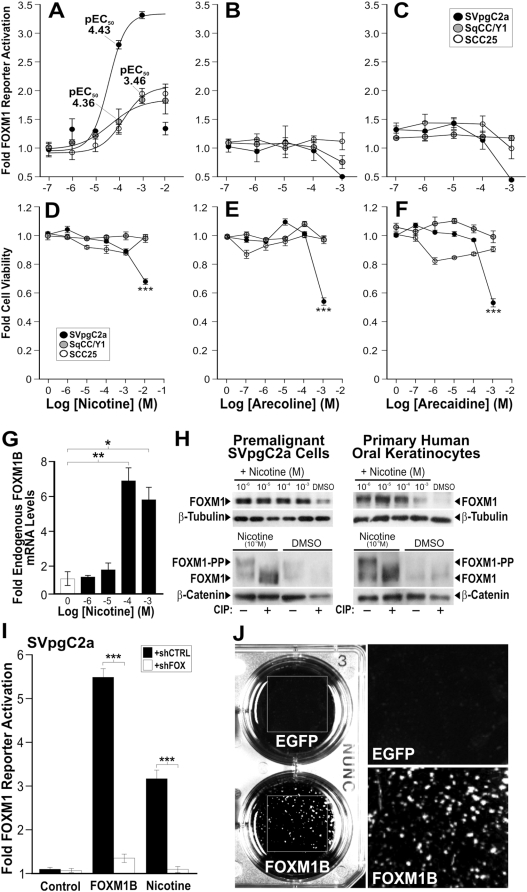
Methodology/principal findings: FOXM1 mRNA and protein expressions were investigated in four independent cohorts (total 75 patients) consisting of normal, premalignant and HNSCC tissues and cells using quantitative PCR (qPCR), expression microarray, immunohistochemistry and immunocytochemistry. Effect of putative oral carcinogens on FOXM1 transcriptional activity was dose-dependently assayed and confirmed using a FOXM1-specific luciferase reporter system, qPCR, immunoblotting and short-hairpin RNA interference. Genome-wide single nucleotide polymorphism (SNP) array was used to 'trace' the genomic instability signature pattern in 8 clonal lines of FOXM1-induced malignant human oral keratinocytes. Furthermore, acute FOXM1 upregulation in primary oral keratinocytes directly induced genomic instability. We have shown for the first time that overexpression of FOXM1 precedes HNSCC malignancy. Screening putative carcinogens in human oral keratinocytes surprisingly showed that nicotine, which is not perceived to be a human carcinogen, directly induced FOXM1 mRNA, protein stabilisation and transcriptional activity at concentrations relevant to tobacco chewers. Importantly, nicotine also augmented FOXM1-induced transformation of human oral keratinocytes. A centrosomal protein CEP55 and a DNA helicase/putative stem cell marker HELLS, both located within a consensus loci (10q23), were found to be novel targets of FOXM1 and their expression correlated tightly with HNSCC progression.

Conclusions/significance: This study cautions the potential co-carcinogenic effect of nicotine in tobacco replacement therapies. We hypothesise that aberrant upregulation of FOXM1 may be inducing genomic instability through a program of malignant transformation involving the activation of CEP55 and HELLS which may facilitate aberrant mitosis and epigenetic modifications. Our finding that FOXM1 is upregulated early during oral cancer progression renders FOXM1 an attractive diagnostic biomarker for early cancer detection and its candidate mechanistic targets, CEP55 and HELLS, as indicators of malignant conversion and progression.


Conflict of interest statement

Competing Interests: The authors have declared that no competing interests exist.

Figures

Figure 1
Figure 1. Upregulation of FOXM1 in both human oral premalignant and HNSCC tissues.
(A) Semi-quantitative RT-PCR showing the relative expression levels of total FOXM1 mRNA in normal oral mucosa (NOM; #1–2), moderate dysplasia (MD; #3–4), severe dysplasia (SD; #5–6), primary HNSCC (#7–8), premalignant oral keratinocytes SVpgC2a, HNSCC cell lines SqCC/Y1 and SCC25. POLR2A was used as an endogenous reference gene. (B) Bioinformatics analysis of microarray data performed on primary cells extracted from normal oral mucosa (NOM), leukoplakia, erythroplakia and primary HNSCC with sample number as indicated. Statistically significant (***P<0.001) activation of FOXM1 mRNA levels when compared to NOM. (C) Quantitative real-time RT-PCR showing relative fold change in FOXM1 mRNA expression levels in HNSCC-derived keratinocytes (n = 15) compared to normal oral mucosa keratinocytes (NOK; n = 4). (D–I) Immunohistochemistry of FOXM1 protein on a panel of FFPE normal oral mucosa (D), mild/moderate dysplasia (E), moderate/severe dysplasia (F), severe dysplasia/carcinoma in situ (G), primary HNSCC (H) and lymph node HNSCC metastasis (I). (J) Digital pixel densitometry for FOXM1 protein immunoreactivity in a panel of 25 oral tissues (n = 5 in each group) as shown in D–I. **(P<0.01) and ***(P<0.001) indicate statistically significant elevation of FOXM1 protein levels when compared to NOM tissues.
Figure 2
Figure 2. Intracellular expression of endogenous FOXM1 protein in primary cultures of human normal oral mucosa (NHOK1), dysplasia (DOK) and HNSCC (UK1).
(A) Fluorescence images showing intracellular localisation of FOXM1 (green) in respective cells counterstained by a nuclear-stain DAPI (blue). The bottom panels are FOXM1 and DAPI merged images of high-power magnification (×400) showing nuclear localisation of FOXM1 protein. (B) Histogram of FOXM1 protein expression levels in 50 cells analysed arranged from low to high FOXM1 levels. (C) Mean FOXM1 protein expression levels in HNOK1, DOK and UK1 cells, n = 50 cells analysed in (B). (D) Immunoblotting for endogenous FOXM1 protein in normal (NHOK1 and NHOK355), dysplasia (POE9n and D20) and HNSCC (CaLH2, CaDec12, UK1 and 5PT) cell lines as indicated. β-Tubulin was immunoblotted for loading control in each sample. (E) Serum starvation (24 h) did not alter the endogenous FOXM1B mRNA levels in primary NHOK1, premalignant POE9n or UK1 HNSCC cells. (F) qPCR data showed that known FOXM1B target genes such as CENPF, CENPA, NEK2 and cyclin B1 are upregulated in UK1 compared to NHOK1 cells. *(P<0.05), **(P<0.01) and ***(P<0.001) indicate significant increase in FOXM1 protein levels when compared to NHOK1 cells.
Figure 3
Figure 3. Upregulation of FOXM1 in various human premalignant and primary cancers.
(A) Bioinformatics analysis of published microarray data on FOXM1 gene expression levels comparing various human tissue types of normal, premalignant and cancers. (B) Fold activation of FOXM1 gene expression in individual tissue types over normal tissues as indicated.
Figure 4
Figure 4. Nicotine activated endogenous FOXM1 transcriptional activity and promoted FOXM1B-induced malignant transformation in human oral keratinocytes.
(A–C) Dose-response curves of nicotine (A), arecoline (B) and arecaidine (C) on FOXM1 transcriptional activity in SVpgC2a, SqCC/Y1 and SCC25 cells, respectively. (D–F) Cell viability assays on SVpgC2a, SqCC/Y1 and SCC25 cells treated with the indicated doses of nicotine (D), arecoline (D) and arecaidine (F), respectively. Each data point indicates mean±SEM (n = 3). ***(P<0.001) indicates significant cell death. (G) qPCR shows that nicotine dose-dependently activated endogenous FOXM1B in SVpgC2a cells. (H) Immunoblotting shows that nicotine dose-dependently activated endogenous FOXM1 protein in both the SVpgC2a (left panel) and primary normal human oral keratinocytes (right panel) as indicated. Bottom panels showed that protein lysates were either pretreated with or without calf-intestinal phosphatase (CIP) prior to immunoblotting. Nicotine at respective optimal doses for each cell type showed reduction of the phosphorylated FOXM1 (FOXM1-PP, top bands) in CIP-treated lysates. β-Tubulin and β-catenin were used as loading control markers. (I) Reversal of nicotine-induced FOXM1 activation by RNA interference shFOX. FOXM1 transcriptional activity was assayed in SVpgC2a cells co-transfected with either shCTRL (control shRNA; solid bars) or FOXM1-specific shFOX (open bars) in either control (mock transfection), FOXM1-overexpressed or nicotine (1 mM)-treated cells, as indicated. ***(P<0.001) indicates significant repression of FOXM1 transcriptional activity over control cells. (J) Anchorage-independent cell transformation assays of EGFP or FOXM1B-overexpressing and nicotine-treated (10 mM) SVpgC2a cells.
Figure 5
Figure 5. Upregulation of FOXM1B induces non-random genomic instability.
(A) A schematic flow diagram showing the approach used in this study to systematically ‘trace’ the genomic instability patterns (LOH and CNA) within the genomes of 8 individual nicotine/FOXM1B-transformed clones using SNP microarrays. Levels of LOH (B), CNA (C), FOXM1B mRNA (using isoform-specific qPCR, see Supplemental Fig. S3) (D) and FOXM1 protein (E) in respective cell clones. (F) Levels of LOH and CNA in primary human normal oral keratinocytes expressing either EGFP or FOXM1B (n = 3). *(P<0.05), **(P<0.01) and ***(P<0.001) indicate significant increase over control cells.
Figure 6
Figure 6. Identification of consensus LOH micro-loci in FOXM1B-transformed SVpgC2a cells.
(A) Alignment of genome-wide LOH profiles of non-transformed (FOXM1B) and FOXM1B-transformed SVpgC2a clones (SVFN1-8). Black lines indicate LOH at a given SNP loci. Histogram (red bars) displays consensus regions of LOH across the 8 SVFN transformed clones. (B) Magnified and detail chromosomal view of the three consensus micro-loci within chromosome 4, 10 and 18 with a list of genes located within each locus as indicated. Uniparental disomy (UPD, blue); copy number gain (CN gain, red); copy number loss (CN loss, green); consensus LOH loci in at least 6 SVFN clones (pink).
Figure 7
Figure 7. CEP55 and HELLS are putative FOXM1B target genes and are upregulated in both premalignant and HNSCC cell lines.
(A) mRNA levels of CEP55 and HELLS but not CENTD1 were significantly upregulated in SVFN transformed clones (solid bars) compared to non-transformed SVpgC2a parental wild-type cells (open bars). (B) Exogenous overexpression of FOXM1B (determined using isoform-specific qPCR, see Supplemental Fig S3), but not MCS (empty vector) or EGFP, in primary human normal oral keratinocytes (NHOK1) showed significant (***P<0.001) induction of endogenous mRNA of CEP55 and HELLS, respectively. Fold mRNA expression levels of FOXM1B (C), CEP55 (D) and HELLS (E) in a panel of oral premalignant cell lines (SVpgC2a, DOK, D19, D20, POE9n-hTERT, POE9n and OKT6) and HNSCC (CA1, UK1, CaLH2, CaLH3, CaDec11, CaDec12, H357, 5PT, PE3/JA, VB6, CaLH2-R, BICR31, SCC4, SCC9, SCC15 and SqCC/Y1) compared to the mean±SEM (n = 8) of normal primary human oral mucosa cells (NHOK1-5, 16, 376, 881). Insets show mean±SEM fold mRNA expression levels of normal (n = 8), oral premalignant (n = 7) and HNSCC cells (n = 16). All three genes show statistically significant (*P<0.05; **P<0.01; ***P<0.001) upregulation in both oral premalignant and HNSCC cell lines. (F) Linear regression analysis between FOXM1B and CEP55 expression in the panel of cell lines used in C and D. (G) Linear regression analysis between FOXM1B and HELLS expression in the panel of cell lines used in C and E. (H) Linear regression analysis between CEP55 and HELLS expression in the panel of cell lines used in D and E. Degree of correlation values (R2) are given in each panel as indicated. (I–J) ChIP-qPCR assays for promoters of CEP55 and HELLS in normal keratinocytes transduced with either Mock (empty virus), EGFP or FOXM1B. Antibodies for GAPDH and cMYC were used as controls for immunoprecipitiation. Representative qPCR curves and melting peaks of each ChIP fractions could be found in Supplemental Fig. S7.
Figure 8
Figure 8. A model mechanism of FOXM1-induced oncogenesis.
(A) FOXM1 expression is upregulated early during the progression from dysplasia to HNSCC malignancy. Nicotine may promote tumour development by synergising with the upregulation of FOXM1 during oncogenesis. (B) Oncogenic 1st hit represents overexpression of FOXM1 which directly activates CEP55 but indirectly activates HELLS expressions. This may destabilise the genome through aberrant mitotic division/cytokinesis and/or epigenetic modification events induced by CEP55 and HELLS, respectively. Oncogenic 2nd hit represents the subsequent accumulation of further genomic instability (for example 10q23 amplification) which promotes oncogenesis.

References

    1. Katoh M. Human FOX gene family (Review). Int J Oncol. 2004;25:1495–1500. - PubMed
    1. Wierstra I, Alves J. FOXM1, a typical proliferation-associated transcription factor. Biol Chem. 2007;388:1257–1274. - PubMed
    1. Korver W, Schilham MW, Moerer P, van den Hoff MJ, Dam K, et al. Uncoupling of S phase and mitosis in cardiomyocytes and hepatocytes lacking the winged-helix transcription factor Trident. Curr Biol. 1998;8:1327–1330. - PubMed
    1. Laoukili J, Kooistra MR, Bras A, Kauw J, Kerkhoven RM, et al. FoxM1 is required for execution of the mitotic programme and chromosome stability. Nat Cell Biol. 2005;7:126–136. - PubMed
    1. Teh MT, Wong ST, Neill GW, Ghali LR, Philpott MP, et al. FOXM1 is a downstream target of Gli1 in basal cell carcinomas. Cancer Res. 2002;62:4773–4780. - PubMed

Publication types

MeSH terms