. 2008:61:1-20.
doi: 10.3114/sim.2008.61.01.

Drought meets acid: three new genera in a dothidealean clade of extremotolerant fungi

Affiliations

Drought meets acid: three new genera in a dothidealean clade of extremotolerant fungi

L Selbmann et al. Stud Mycol. 2008.

Abstract

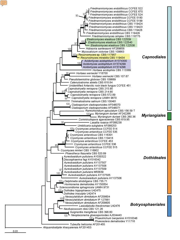
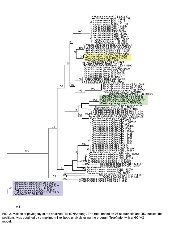
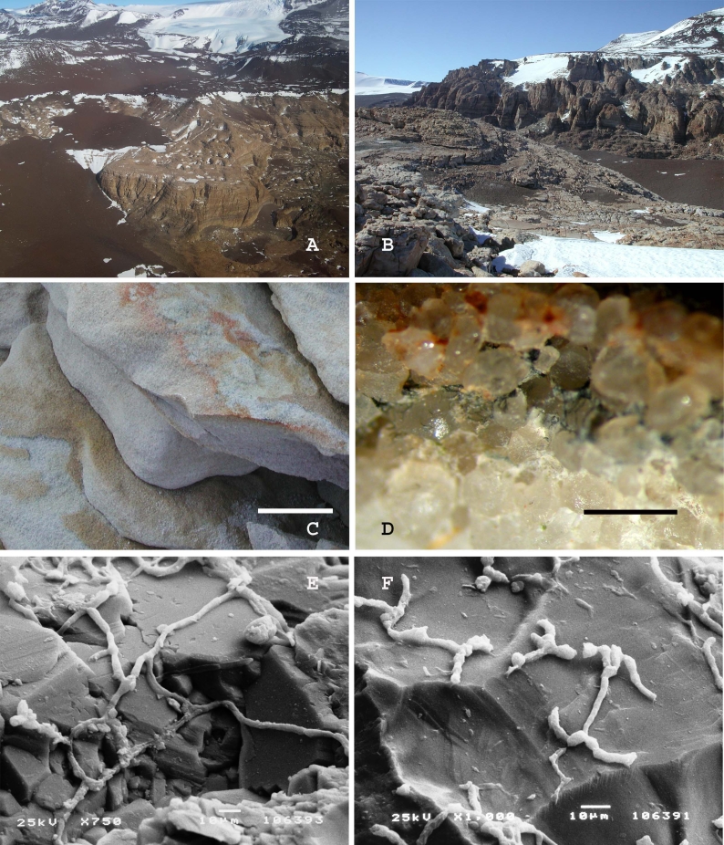
Fungal strains isolated from rocks and lichens collected in the Antarctic ice-free area of the Victoria Land, one of the coldest and driest habitats on earth, were found in two phylogenetically isolated positions within the subclass Dothideomycetidae. They are here reported as new genera and species, Recurvomyces mirabilisgen. nov., sp. nov. and Elasticomyces elasticusgen. nov., sp. nov. The nearest neighbours within the clades were other rock-inhabiting fungi from dry environments, either cold or hot. Plant-associated Mycosphaerella-like species, known as invaders of leathery leaves in semi-arid climates, are also phylogenetically related with the new taxa. The clusters are also related to the halophilic species Hortaea werneckii, as well as to acidophilic fungi. One of the latter, able to grow at pH 0, is Scytalidium acidophilum, which is ascribed here to the newly validated genus Acidomyces. The ecological implications of this finding are discussed.

Keywords: Acidophilic fungi; Antarctica; ITS; SSU; black fungi; extremotolerance; halophilic fungi; lichens; phylogeny; rock-inhabiting fungi; taxonomy.


Figures

Fig. 1.
Fig. 1.
Molecular phylogeny based on SSU sequences indicating the positions of the clades in Dothideomycetidae; the described new genera were highlighted with coloured rectangles. The tree has been built with neighbour-joining algorithm in ARB package with 100 replications. Branches of the clades supported by a bootstrap value above 95 % are in bold.
Fig. 2.
Fig. 2.
Molecular phylogeny of the analysed ITS rDNAs showing the relationship of the new genera described, highlighted with coloured rectangles, in the Capnodiales. The neighbour-joining tree, based on 95 sequences and 452 nucleotide positions, has been generated using HKY+G model; the model was calculated using ML in MrAIC software. Bootstrap values from 100 resampled data sets are shown.
Fig. 3.
Fig. 3.
Recurvomyces mirabilis. A. 1- celled conidia and ramoconidia. B–D. Septate and branched conidiophores; branches forming right angle and bent down. E. Conidiophore with recurved hyphal branching. F. Conidiogenous cells producing conidia by enteroblastic proliferation. G. Schyzolytic conidial secession. H. Swelling cells. Scale bar = 10 μm.
Fig. 4.
Fig. 4.
A. Battleship Promontory, Southern Victoria Land, Antarctica, view from the helicopter. B. Battleship Promontory, Southern Victoria Land, Antarctica, landscape. C. Sandstone sample from which Recurvomyces mirabilis CBS 119434 has been isolated. The surface is weathered by the activity of cryptoendolithic microorganisms. Scale bar = 5 cm. D. Magnification at the dissecting microscope of the black fungi endolithic colonization inside the sample of sandstone used for the isolation. Scale bar = 2 mm. E, F. Rock colonised by the fungus observed at the Scanning Electron Microscope.
Fig. 5.
Fig. 5.
Recurvomyces mirabilis, CBS 119434 (= CCFEE 5264). A. Strain grown on different media after two mo of incubation at 15 °C. B–D. Hyphae with branched and unbranched conidiophores producing 0–1 septated conidia. E. Curved branched conidiophores schyzolytically seceding (black arrow) showing enteroblastic elongation (white arrow) at the apex. F, G. high magnification of branched conidiophores producing 1-celled conidia. H, I. 1- and 2-celled conidia and ramoconidia. L. Terminal swelling cell. M, N. swelling hyphae showing enteroblastic elongation. O, P. Unbranched conidiophores producing 1-celled conidia, scar is visible after schizolytic secession (P, arrow). Q. Branched conidiophore. R, S. swelling cell at intercalary (R) and terminal position (S). B–N. Light microscopy; Scale bars = 10 μm. O–S. SEM.
Fig. 6.
Fig. 6.
Elasticomyces elasticus. A. Vegetative hyphae. B, C. Branched fertile hyphae producing conidia by fragmentation. D. Disarticulating fertile hyphae where cells remain joint by connectives. E. Swelling hyphae. Scale bar = 20 μm.
Fig. 7.
Fig. 7.
Elasticomyces elasticus, CCFEE 5313. A. Strain grown on different media after 1.5 mo of incubation at 15 °C. B–D. Vegetative and fertile hyphae. E–H. High magnification of fertile hyphae; anastomoses in the vegetative hyphae (black arrows in E). I–O. Uncompleted disarticulation of artic conidia and hyphal fragments remaining joint by connectives (white arrows). P. High magnification of conidia remaining joint after disarticulation. Q, R. 1- and 2-celled conidia produced after schyzolithic secession (white arrow). S–U. Enteroblastic elongation at the apexes (white arrows). Scale bars = 20 μm.
Fig. 8.
Fig. 8.
Acidomyces acidophilus. A. Strain CBS 899.87 grown on different media after 1 mo of incubation at 25 °C. B–D, G. Toruloid unbranched hyphae with melanised and thick-walled cells. E. Meristematic development of the hyphae. F. Fungus grown at pH 1 in liquid culture. H. Chain of 1- 2- and 3- celled conidia. I. Strain CBS 335.97 grown on different media after 1 mo of incubation at 25 °C. L, M. Filamentous hyphae with intercalary and terminal swelling cells (black arrows). N. Fungus grown at pH 1 in liquid culture. Scale bars = 20 μm.

References

    1. Baker BJ, Lutz MA, Dawson SC, Bond PL, Banfield JF (2004). Metabolically active eukaryotic communities in extremely acidic mine drainage. Applied and Environmental Microbiology 70: 6264–6271. - PMC - PubMed
    1. Bills GF, Collado J, Ruibal C, Peláez F, Platas G (2004). Hormonema carpetanum sp. nov., a new lineage of dothideaceous black yeasts from Spain. Studies in Mycology 50: 149–157.
    1. Bogomolova EV, Minter DW (2003). A new microcolonial rock-inhabiting fungus from marble in Chersonesos (Crimea, Ukraine). Mycotaxon 86: 195–204.
    1. Bruns TD, Vilgalys R, Barns SM, Gonzalez D, Hibbett DS, Lane DJ, Simon L, Stickel S, Szaro TM, Weisburg WG (1992). Evolutionary relationships within the fungi: analyses of nuclear small subunit rRNA sequences. Molecular Phylogenetics and Evolution 1: 231–241. - PubMed
    1. Butin H, Pehl L, Hoog GS de, Wollenzien U (1996). Trimmatostroma abietis sp. nov. (Hyphomycetes) and related species. Antonie van Leeuwenhoek 69: 203–209. - PubMed

LinkOut - more resources