. 2008:61:175-91.
doi: 10.3114/sim.2008.61.18.

Biodiversity of the genus Cladophialophora

Affiliations

Biodiversity of the genus Cladophialophora

H Badali et al. Stud Mycol. 2008.

Abstract

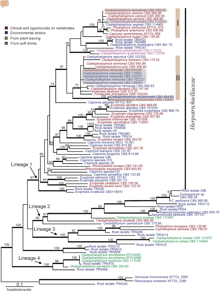
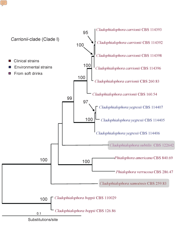
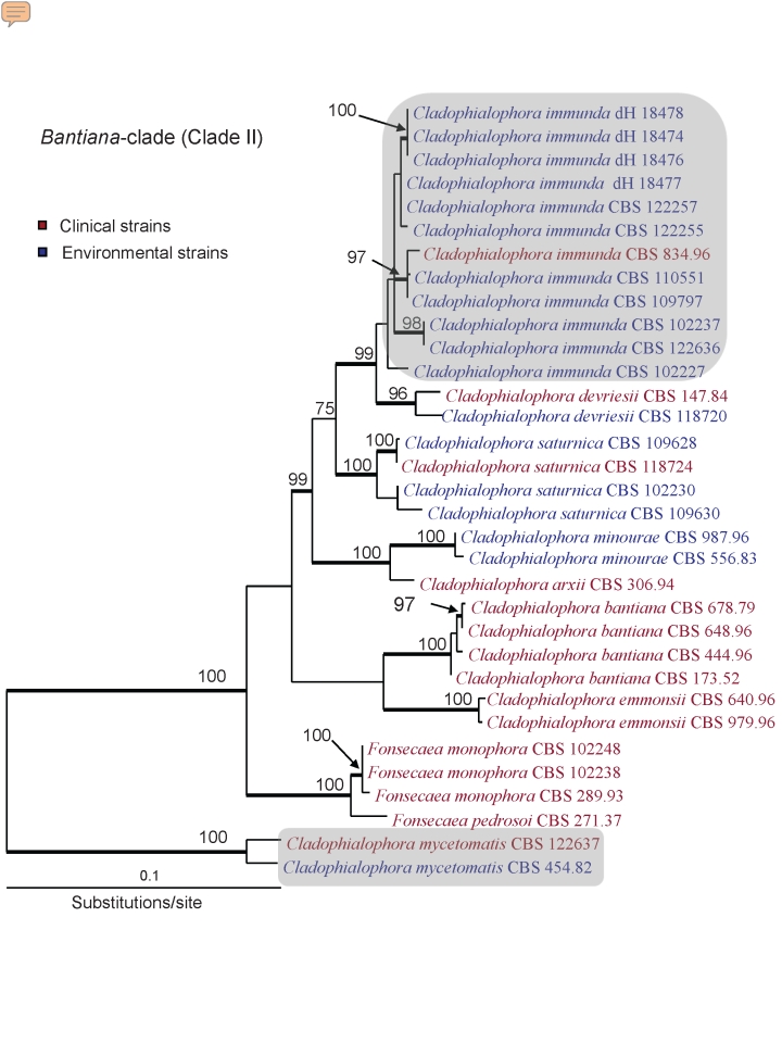
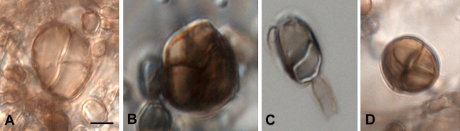
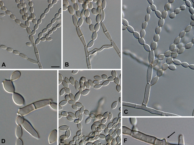
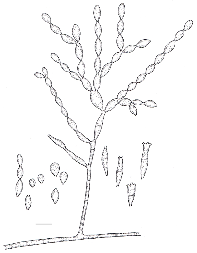
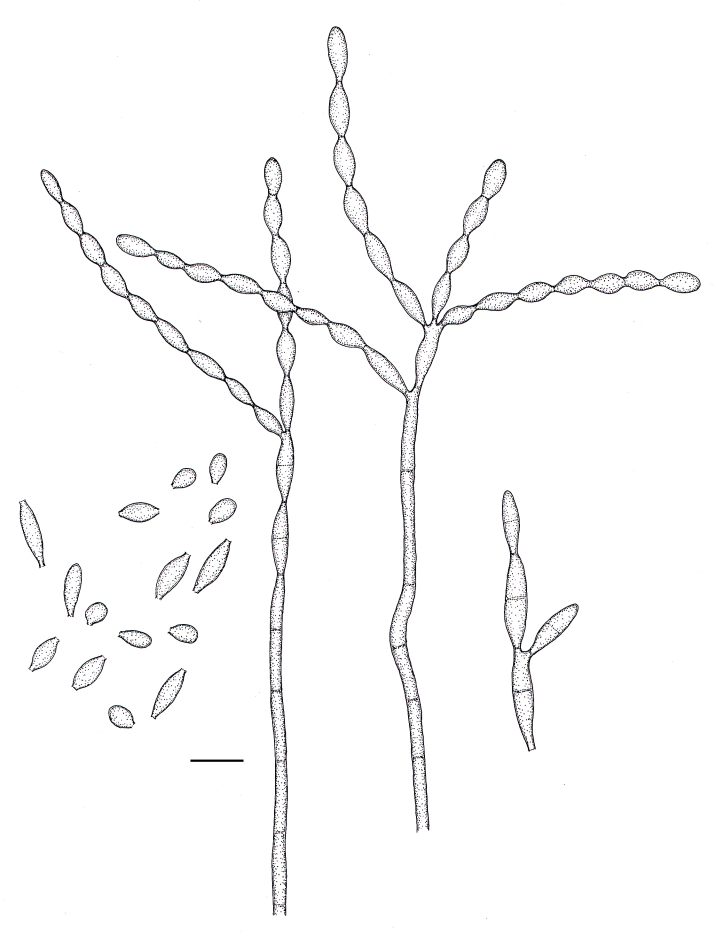
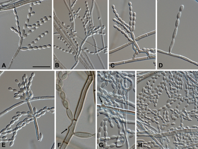
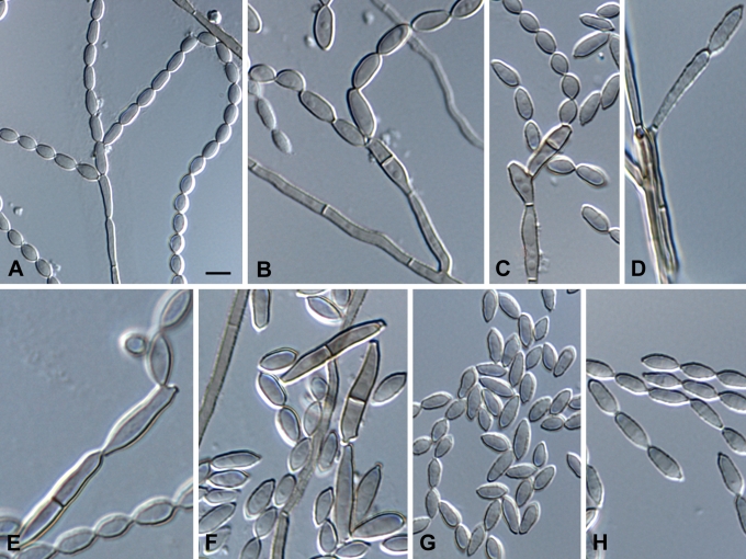
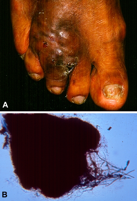
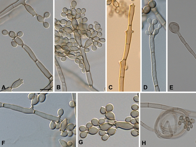
Cladophialophora is a genus of black yeast-like fungi comprising a number of clinically highly significant species in addition to environmental taxa. The genus has previously been characterized by branched chains of ellipsoidal to fusiform conidia. However, this character was shown to have evolved several times independently in the order Chaetothyriales. On the basis of a multigene phylogeny (nucLSU, nucSSU, RPB1), most of the species of Cladophialophora (including its generic type C. carrionii) belong to a monophyletic group comprising two main clades (carrionii- and bantiana-clades). The genus includes species causing chromoblastomycosis and other skin infections, as well as disseminated and cerebral infections, often in immunocompetent individuals. In the present study, multilocus phylogenetic analyses were combined to a morphological study to characterize phenetically similar Cladophialophora strains. Sequences of the ITS region, partial Translation Elongation Factor 1-alpha and beta-Tubulin genes were analysed for a set of 48 strains. Four novel species were discovered, originating from soft drinks, alkylbenzene-polluted soil, and infected patients. Membership of the both carrionii and bantiana clades might be indicative of potential virulence to humans.

Keywords: Biodiversity; Cladophialophora; MLST; bioremediation; chromoblastomycosis; disseminated infection; mycetoma.


Figures

Fig. 1.
Fig. 1.
Phylogeny obtained from a ML analysis of three combined loci (SSU, LSU and RPB1) using RAxML. Bootstrap support values were estimated based on 500 replicates, and are shown above the branches (thick branch for values ≥ 70 %). The tree was rooted using Verrucula inconnexaria, Placocarpus schaereri and the rock isolate TRN242. New species are highlighted with grey boxes.
Fig. 2.
Fig. 2.
Phylogeny of Clade I obtained from a ML analysis of three combined loci (ITS rDNA, EF1-α and TUB) using RAxML. Bootstrap support values were estimated based on 500 replicates, and are shown above the branches (thick branch for values ≥ 70 %). New species are highlighted using with grey boxes. Cladophialophora boppii (CBS 126.86 and CBS 110029) were taken as outgroup.
Fig. 3.
Fig. 3.
Phylogeny of Clade II obtained from a ML analysis of three combined loci (ITS rDNA, EF1-α and TUB) using RAxML. Bootstrap support values were estimated based on 500 replicates, and are shown above the branches (thick branch for values ≥ 70 %). New species are highlighted using with grey boxes. The tree was rooted with two strains of Cladophialophora mycetomatis (CBS 454.82 and CBS 122637).
Fig. 4.
Fig. 4.
Colony diameters of novel Cladophialophora species at different temperatures ranging from 6 to 40 °C, measured after two wks on 2 % MEA.
Fig. 5.
Fig. 5.
Morphology of muriform cells in three species of Cladophialophora. A–B. Cladophialophora subtilis. C. Cladophialophora immunda. D. Cladophialophora carrionii. Scale bar = 10 μm.
Fig. 6.
Fig. 6.
Cladophialophora samoënsis (CBS 259.83). A. Conidiophore. B–E. Conidial chains with ramoconidia and conidia. F. Conidiogenous loci (arrows). Scale bar = 10 μm.
Fig. 7.
Fig. 7.
Microscopic morphology of C. samoënsis (CBS 259.83). Branched conidial chains with ramoconidia and conidia. Conidiophores septate, lateral or terminal, with denticles on the stipe and on 0–1-septate ramoconidia. Scale bar = 10 μm.
Fig. 8.
Fig. 8.
Microscopic morphology of C. subtilis (CBS 122642). Fertile hyphae septate, ascending to erect. Conidiophores apically branched, cylindrical to sub-cylindrical Branched conidial chains with ramoconidia and conidia. Scale bar = 10 μm.
Fig. 9.
Fig. 9.
Microscopic morphology of C. mycetomatis (CBS 122637 and CBS 454.82). Septate hyphae creeping, ascending to sub-erect. Conidiophores solitary, micronematous, cylindrical, apically branched. Conidia holoblastic, fusiform produced in long chains. Scale bar = 10 μm.
Fig. 10.
Fig. 10.
Microscopic morphology of C. immunda (CBS 834.96, CBS 109797, CBS 110551, CBS 102227, CBS 102237). Hyphae branched, septate, straight, ascending to erect, Hyphae giving rise to conidiophores. Lemon-shaped to pyriform to guttuliform, narrowed towards one or both ends, coherent or deciduous. Scale bar = 10 μm.
Fig. 11.
Fig. 11.
Cladophialophora subtilis (CBS 122642). A–D. Conidiophores with branched conidial chains and ramoconidia. E. Sympodial conidiogenesis. F–G. Conidiogenous loci (arrows). H. Conidia. Scale bars = 10 μm.
Fig. 12.
Fig. 12.
Cladophialophora mycetomatis (CBS 122637 and CBS 454.82). A–B. Conidiophores and conidial chains with ramoconidia and conidia. C–D. Cylindrical, septate conidiophores. G. Ellipsoidal to fusiform conidia. Scale bars = 10 μm.
Fig. 13.
Fig. 13.
Clinical manifestations of Eumycetoma caused by Cladophialophora mycetomatis. A. Deformed tumorous area of the foot, with nodules, draining sinuses and discharging purulent fluid. B. Branched black granule, approximately 500 μm in size (in 10 % KOH), with septate hyphae.
Fig. 14.
Fig. 14.
Cladophialophora immunda (CBS 834.96, CBS 109797, CBS 110551, CBS 102227, CBS 102237). A–D. Conidiophores and conidial apparatus with T-shaped foot cell and cylindrical, septate, denticulate conidiogenesis. E. Chlamydospores. F–G. Thin-walled, lemon-shaped to pyriform to guttuliform conidia. H. Hyphal coil. Scale bar = 10 μm.
Fig. 15.
Fig. 15.
Cladophialophora immunda (CBS 834.96). A. Gomori Methenamine-silver and B. Periodic acid-Schiff stained sections revealed septate hyphae, moniliform hyphae of different lengths, and thick-walled cells. (GMS & PAS × 360)

References

    1. Badali H, Carvalho VO, Vicente V, Attili-Angelis D, Kwiatkowski IB, Gerrits van den Ende AHG, Hoog GS de (2008). Cladophialophora saturnica sp. nov., a new opportunistic species of Chaetothyriales revealed using molecular data. Medical Mycology 7: 1–12. - PubMed
    1. Borelli D (1980). Causal agents of chromoblastomycosis (Chromomycetes). Proceedings of the 5th International Conference on Mycoses pp. 335–340.
    1. Braun U (1998). A monograph of Cercosporella, Ramularia and allied genera(Phytopathogenic Hyphomycetes). Vol. 2. IHW-Verlag, Eching.
    1. Braun U, Crous PW, Dugan F, Groenewald JZ, Hoog GS de (2003). Phylogeny and taxonomy of Cladosporium-like hyphomycetes, including Davidiella gen. nov., the teleomorph of Cladosporium s. str. Mycological Progress 2: 3–18.
    1. Braun U, Feiler U (1995). Cladophialophora and its teleomorph. Microbiological Research 150: 81–91.

LinkOut - more resources