Comparative Study
. 2009 Jun;65(6):667-76.
doi: 10.1002/ana.21627.

Efficacy of systemic morpholino exon-skipping in Duchenne dystrophy dogs

Affiliations
Comparative Study

Efficacy of systemic morpholino exon-skipping in Duchenne dystrophy dogs

Toshifumi Yokota et al. Ann Neurol. 2009 Jun.

Abstract

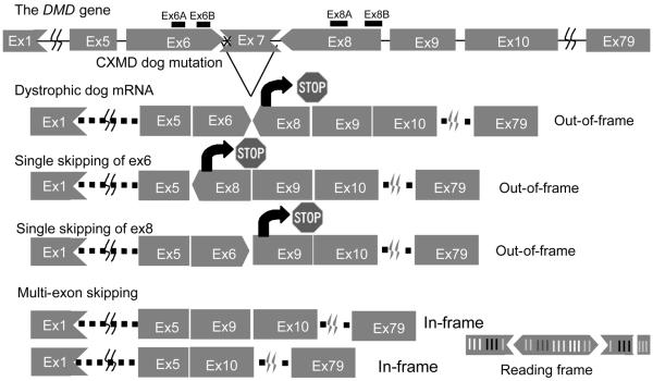
Objective: Duchenne muscular dystrophy (DMD) is caused by the inability to produce dystrophin protein at the myofiber membrane. A method to rescue dystrophin production by antisense oligonucleotides, termed exon-skipping, has been reported for the mdx mouse and in four DMD patients by local intramuscular injection. We sought to test efficacy and toxicity of intravenous oligonucleotide (morpholino)-induced exon skipping in the DMD dog model.

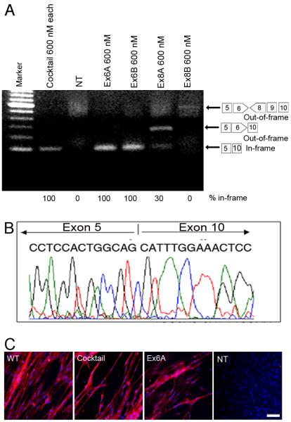
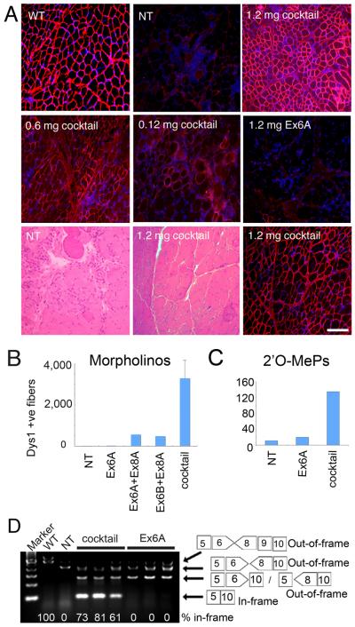
Methods: We tested a series of antisense drugs singly and as cocktails, both in primary cell culture, and two in vivo delivery methods (intramuscular injection and systemic intravenous injection). The efficiency and efficacy of multiexon skipping (exons 6-9) were tested at the messenger RNA, protein, histological, and clinical levels.

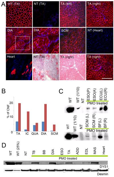
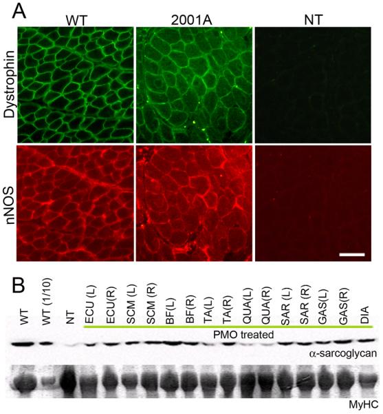
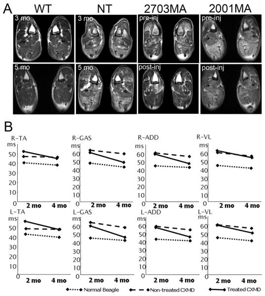
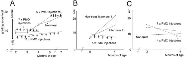
Results: Weekly or biweekly systemic intravenous injections with a three-morpholino cocktail over the course of 5 to 22 weeks induced therapeutic levels of dystrophin expression throughout the body, with an average of about 26% normal levels. This was accompanied by reduced inflammatory signals examined by magnetic resonance imaging and histology, improved or stabilized timed running tests, and clinical symptoms. Blood tests indicated no evidence of toxicity.

Interpretation: This is the first report of widespread rescue of dystrophin expression to therapeutic levels in the dog model of DMD. This study also provides a proof of concept for systemic multiexon-skipping therapy. Use of cocktails of morpholino, as shown here, allows broader application of this approach to a greater proportion of DMD patients (90%) and also offers the prospect of selecting deletions that optimize the functionality of the dystrophin protein.


Figures

Figure 1
Figure 1. Mutation in CXMD and strategy for exon skipping treatment
The dystrophic dog harbors a point mutation at splice site in intron 6, which leads to lack of exon 7 in mRNA. Single exon skipping of exon 6 or exon 8 leads to out of frame products. Exclusion of at least two further exons (exon 6 and exon 8) is required to restore reading frame. 1) Vertical line means that the exons begin by the first nucleotide of a codon, 2) Arrows toward left mean that the exons begin with the second nucleotide of a codon, 3) Arrows toward right mean that the exon begin with the third nucleotide of a codon.
Figure 2
Figure 2. In vitro screening of antisense oligonucleotides and recovery of dystrophin expression by single antisense oligos in dog primary myoblasts
(A) Detection of exon 6-9 skipped in-frame products (101 bp) using RT-PCR at 4 days after the transduction of 5 μg (600 nM) each AOs of single (Ex6A, or Ex6B) or cocktail AOs (Ex6A, Ex6B, Ex8A, and Ex8B) as indicated. The faint 585 bp out-of-frame band is detected in Ex8B treated myotubes. Non-treated myotubes (NT) show little RT-PCR product, likely due to nonsense-mediated decay. (B) cDNA sequencing after antisense oligonucleotide treatment at 4 days after the transduction of Ex6A alone, showing the desired in-frame exon 5-10 skip. (C) Immunocytochemistry with dystrophin C-terminal antibody (Dys-2; Red) and nuclear counter staining (Blue) for primary myotubes from CXMD cells at 4 days after transfection with cocktail or single antisense 2'O-MePs targeting exon 6 and 8 (5 μg each/ml, or 600 nM), non-treated wild-type (WT) and CXMD cells (CXMD). Bar; 50 μm.
Figure 3
Figure 3. Recovery of dystrophin expression by in vivo intramuscular injection of a three-AO cocktail, but not Ex6A alone
(A) Restoration of dystrophin expression in TA at 14 days after single injection with 1.2 mg of Ex6A only, or cocktail containing 0.12 mg each, 0.6 mg each, or 1.2 mg each of antisense morpholino Ex6A, Ex6B, and Ex8A are shown. Age-matched non-treated CXMD (NT) and wild-type (WT) dogs are shown as controls. HE staining at 14 days after 1.2 mg each of the cocktail injection and age-matched non-treated control (NT) with consecutive cryosection stained with DYS-1 and DAPI show histological correction of the dystrophy. Bar; 100 μm. (B) and (C) show the number of dystrophin (DYS1) positive fibers in TA or extensor carpi ulnaris at 14 days after a single injection with cocktail (Ex6A+Ex6B+Ex8A), or indicated combinations, at 1.2 mg each (morpholino; B), or 120 μg each 2'O-MePs (C). Values are mean ± s. e. m. (D) RT-PCR analysis at 2 weeks after intramuscular injection of cocktail or Ex6A morpholino at 1.2 mg each. The percentage of the in-frame exons 5-10 skip is shown under the gel image for treated muscle; normal control (WT) muscle shows the normal full-length in-frame transcript at the expected 100%.
Figure 4
Figure 4. Wide-spread dystrophin expression and improved histology by intravenous systemic delivery of cocktail morpholinos in CXMD dogs
(A) Dystrophin (DYS1) staining and histology in bilateral tibialis anterior muscles (TA), diaphragm (DIA), sternocleidomastoid (SCM) and heart at 2 weeks after final injection after 5 × weekly iv injection of 120 mg/kg of cocktail morpholinos containing Ex6A, Ex6B, and Ex8A (2001MA). Comparisons were made with TA from normal control (wild-type; WT) and from non-treated CXMD littermate (NT) tibialis anterior (TA) and heart. Intravenous morpholino treatment resulted in extensive though variable dystrophin production in multiple muscles, but with only limited evidence of rescue in heart (isolated cardiocytes). Paired dystrophin immunostaining and histology from treated dog (TA, lower panels) showed improved histopathology relative to untreated littermate (NT TA) histology. Bars; 200 μm, except for higher magnification picture of DIA and hearts (100 μm). (B) Quantitation of centrally nucleated fibers (CNFs) in TA, intercostal (IC), Quadriceps (QUA), diaphragm (DIA), and sternocleidomastoid (SCM) in treated dog (blue bars; 2001MA) and untreated dog (red bars; 2008MA). (C) Western blotting analysis for detection of dystrophin at 2 weeks after final injection after 5 × weekly iv injection of 120 mg/kg of cocktail morpholinos containing Ex6A, Ex6B, and Ex8A (2001MA). Dystrophin rescue is variable with high expression in right extensor carpi ulnaris (ECU(R)) and left biceps femoris (BF(L)), and less in posterior or anterior esophagus (ESO(P), ESO(A)), and sternocleidomastoid (SCM). (D) Immunoblot analysis of dystrophin in intravenous morpholino treated dog (2703MA; 7× weekly dosing) and controls (normal control [WT], non-treated [NT]). Desmin immunoblot is shown as a loading control. Dystrophin shows high levels (>25% control levels) in triceps brachii (TB), diaphragm (DIA) and masseter (MAS). ESO Esophagus (posterior or anterior); ECU, Extensor carpi ulnaris; SCM, Sternocleidomastoid; BF, Biceps Femoris; TB, Triceps Brachii; BB, Biceps Brachii; DIA, Diaphragm; ADD, Adductor; EDL, Extensor digitorum longus; MAS, Masseter.
Figure 5
Figure 5. Recovery of localization and expression of dystrophin associated proteins after systemic delivery of cocktail morpholinos to CXMD dogs
nNOS (A) and α-sarcoglycan (B) expression at 2 weeks after 5 × weekly 120 mg/kg cocktail (2001MA) or 7 × weekly 200 mg/kg cocktail morpholino injections (2703MA) CXMD dogs. Recovery of nNOS expression at sarcolemma was observed by double immuno-fluorescence against dystrophin (DYS-1) and nNOS. Bar; 50 μm. By immunoblot (B), α-sarcoglycan levels are increased in treated dog muscles, compared to untreated dystrophic controls (NT). Myosin heavy chain (MyHC) shown as a loading control. WT; wild-type normal controls, WT(1/10); wild-type (1/10 diluted samples, i.e. 4 μg loaded), NT; non-treated CXMD muscles (tibialis anterior) ECU; Extensor carpi ulnaris, SCM; Sternocleidomastoid, BF; Biceps Femoris, TA; Tibialis Anterior, QUA; Quadriceps, SAR; Sartorius, GAS; Gastrocnemius, DIA; Diaphragm. (L) and (R) stand for left side and right side respectively.
Figure 6
Figure 6. Amelioration of pathology and reduced inflammation signal in MRI
T2-weighted MRI of hind legs at 1 week before initial injection (pre-inj), and at 2 weeks after final injection (post-inj) of 7 × weekly iv injection of 200 mg/kg of cocktail morpholinos (2703MA) or 5 × weekly iv injection of 120 mg/kg of cocktail morpholinos (2001MA). Age-matched un-treated dogs (WT [normal control] and NT [non-treated dystrophic control]) are shown for comparison. (B) Changes of T2 value examined by MRI at 2 weeks after 7 × weekly 200 mg/kg of cocktail morpholino injections. Changes of T2 values in hind-legs at 1 week before initial injection and at 2 weeks after final injection are shown. Intravenous morpholino treatment resulted in decreased T2 signal in all muscles examined. TA, Tibialis Anterior; GAS, Gastrocnemius; VL, Vastus lateralis; ADD, Adductor.
Figure 7
Figure 7. Stabilization of clinical symptoms by systemic morpholino treatment
(A) A combined clinical grading scores before (black lines) and after starting treatment (red lines) of the three treated dogs. Clinical grades of gait disturbance, mobility disturbance, limb or temporal muscle atrophy, drooling, macroglossia, Dysphagia are scored as described in methods. A series of untreated dogs (n=6-13) were studied for comparison (dashed line, standard error bars). (B–C) Fifteen meter timed running tests in treated dogs and untreated littermates. A CXMD dog treated from 5–7 months (2001MA) of age showed decreased timed 15 m run after treatment, whereas untreated littermates showed slowed running ability (B). Similarly, two littermate dogs treated at 2–4 months of age (2703MA; 2702FA) showed quicker 15 m times following treatment compared to non-treated littermate (C).

References

    1. Hoffman EP, Brown RH, Jr., Kunkel LM. Dystrophin: the protein product of the Duchenne muscular dystrophy locus. Cell. 1987;51:919–928. - PubMed
    1. Matsumura K, Campbell KP. Dystrophin-glycoprotein complex: its role in the molecular pathogenesis of muscular dystrophies. Muscle Nerve. 1994;17:2–15. - PubMed
    1. Koenig M, Beggs AH, Moyer M, et al. The molecular basis for Duchenne versus Becker muscular dystrophy: correlation of severity with type of deletion. Am J Hum Genet. 1989;45:498–506. - PMC - PubMed
    1. England SB, Nicholson LV, Johnson MA, et al. Very mild muscular dystrophy associated with the deletion of 46% of dystrophin. Nature. 1990;343:180–182. - PubMed
    1. Dunckley MGMM, Villiet P, Eperon IC, Dickson G. Modification of splicing in the dystrophin gene in cultured Mdx muscle cells by antisense oligoribonucleotides. Hum Mol Genet. 1998;7:1083–1090. - PubMed

Publication types