. 2009 Apr;9(7):1869-82.
doi: 10.1002/pmic.200701071.

Pharmacologic intervention targeting glycolytic-related pathways protects against retinal injury due to ischemia and reperfusion

Affiliations

Pharmacologic intervention targeting glycolytic-related pathways protects against retinal injury due to ischemia and reperfusion

Ling Zheng et al. Proteomics. 2009 Apr.

Abstract

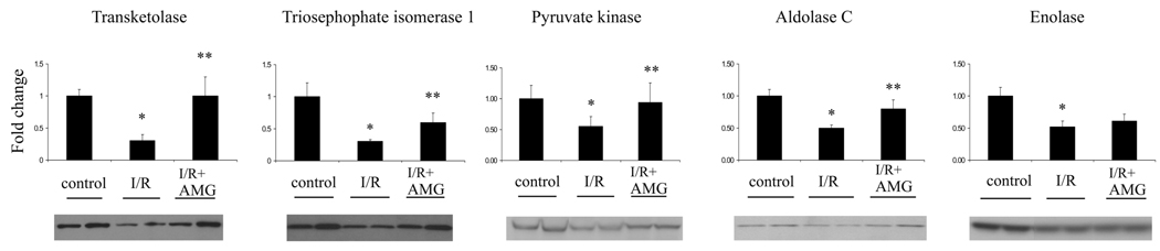
Retinal ischemia contributes to multiple ocular diseases while aminoguanidine (AMG) treatment significantly inhibits the neuronal and vascular degeneration due to acute retinal ischemia and reperfusion (I/R) injury. In the present study, 2-D DIGE was applied to profile global protein expression changes due to retinal I/R injury, and the protection effects mediated by AMG. Retinal ischemia was induced by elevated intraocular pressure to 80-90 mmHg for 2 h, and reperfusion was established afterward. Retinal tissues were collected 2 days after I/R injury. After 2-D DIGE analysis, a total of 96 proteins were identified. Among them, 28 proteins were identified within gel spots whose intensities were normalized by AMG pretreatment, pathway analysis indicated that most were involved in glycolysis and carbohydrate metabolism. Selected enzymes identified by MS/MS within these pathways, including transketolase, triosephosphate isomerase 1, aldolase C, total enolase, and pyruvate kinase were validated by quantitative Western blots. Glycolytic enzymes and other differentially regulated proteins likely play previously unrecognized roles in retinal degeneration after I/R injury, and inhibition of the resulting metabolic changes, using pharmacologically agents such as AMG, serve to inhibit the changes in metabolism and mitigate retinal degeneration. Select glycolytic enzymes may provide novel therapeutic targets for inhibiting the neuronal and vascular degeneration after retinal I/R injury.


Figures

Figure 1
Figure 1
2D gel migration patterns of proteins that normalized by AMG treatment after retinal I/R injury. Cy dye image form samples run on pH 3–10 nonlinear gradient IPG strips and 12% polyacrylamide gel is shown. Numbers in the figure are master spot number that used to match every gel, and spot areas are highlighted. Refer to table 1 for fold changes in expression that calculated using DeCyder software.
Figure 2
Figure 2
Identified, differentially expressed proteins that normalized by AMG treatment after retinal I/R injury were shown by DeCyder output. (A) AMG treatment bring back the protein levels that were decreased after retinal I/R (B) AMG treatment inhibit retinal I/R-induced some proteins’ overexpression. Values on the y-axis are the relative arbitrary units. Circles indicate the value of individual samples. The crosses indicate the average values of each group. (C: control retinas; I/R: ischemia/reperfusion injured retinas; I/R+AMG: AMG pre-treated ischemia/reperfusion injured retinas)
Figure 3
Figure 3
2D gel migration patterns of additional proteins that altered after retinal I/R injury. Cy dye image form samples run on pH 3–10 nonlinear gradient IPG strips and 12% polyacrylamide gel is shown. Numbers in the figure are master spot number, and spot areas are highlighted. Refer to table 2 for fold changes in expression that calculated using DeCyder software.
Figure 4
Figure 4
PANTHER classification by pathway (A), biological process (B) and molecular function (C). (Heterotrimeric G-protein signaling pathway-1* is Gi alpha and Gs alpha mediated; Heterotrimeric G-protein signaling pathway-2* is Gq alpha and Go alpha mediated; Heterotrimeric G-protein signaling pathway-3* involves in rod outer segment phototransduction)
Figure 5
Figure 5
Western blots of several glycolytic enzymes with densitomertric quantitation results on top and representative protein bands at the bottom. Expression is reported relative to β-actin in same sample, and normalized to control retinas, set as 1 fold. (C: control retinas, I/R: ischemia/reperfusion injured retinas; I/R+AMG: AMG pre-treated ischemia/reperfusion injured retinas; * p < 0.05 compared to control retinas; ** p < 0.05 compared to I/R injured retinas)
Figure 6
Figure 6
Alteration of glycolysis and pathways that related to glycolysis after retinal I/R injury. Bold; decreased expression after retinal I/R injury. Italics; increased expression after retinal I/R injury. (LDH, lactate dehydrogenase; IDH, isocitrate dehydrogenase; MDH, malate dehydrogenase)

Similar articles

Cited by

References

    1. Lefer DJ, Granger DN. Am J Med. 2000;109:315–323. - PubMed
    1. Chen JK, Chow SE. Chang Gung Med J. 2005;28:369–377. - PubMed
    1. Kutala VK, Khan M, Angelos MG, Kuppusamy P. Antioxid Redox Signal. 2007;9:1193–1206. - PubMed
    1. Osborne NN, Casson RJ, Wood JP, Chidlow G, et al. Prog Retin Eye Res. 2004;23:91–147. - PubMed
    1. Zheng L, Gong B, Hatala DA, Kern TS. Invest Ophthalmol Vis Sci. 2007;48:361–367. - PubMed

Publication types