. 2009 Sep;76(5):334-44.
doi: 10.1002/cyto.b.20473.

A rapid flow cytometric screening test for X-linked lymphoproliferative disease due to XIAP deficiency

Affiliations

A rapid flow cytometric screening test for X-linked lymphoproliferative disease due to XIAP deficiency

Rebecca A Marsh et al. Cytometry B Clin Cytom. 2009 Sep.

Abstract

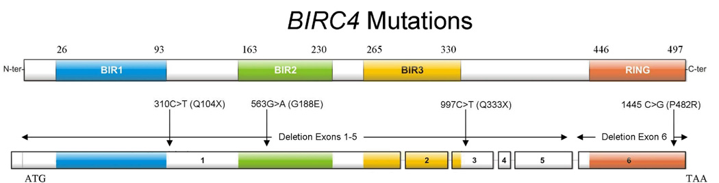
Background: Deficiency of X-linked inhibitor of apoptosis (XIAP), caused by BIRC4 gene mutations, is the second known cause of X-linked lymphoproliferative disease (XLP), a rare primary immunodeficiency that often presents with life-threatening hemophagocytic lymphohistiocytosis (HLH). Rapid diagnosis of the known genetic causes of HLH, including XIAP deficiency, facilitates the initiation of life-saving treatment and preparation for allogeneic hematopoietic cell transplantation (HCT). Until now, a rapid screening test for XIAP deficiency has not been available.

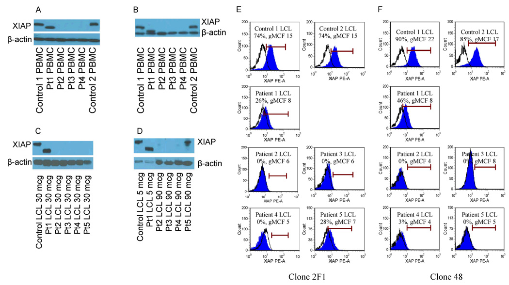
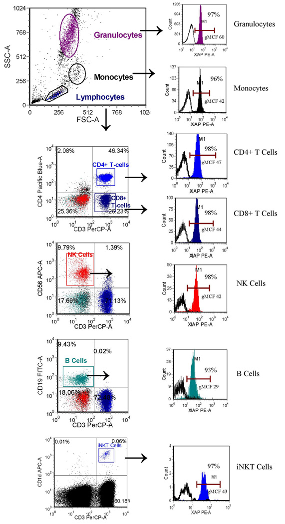
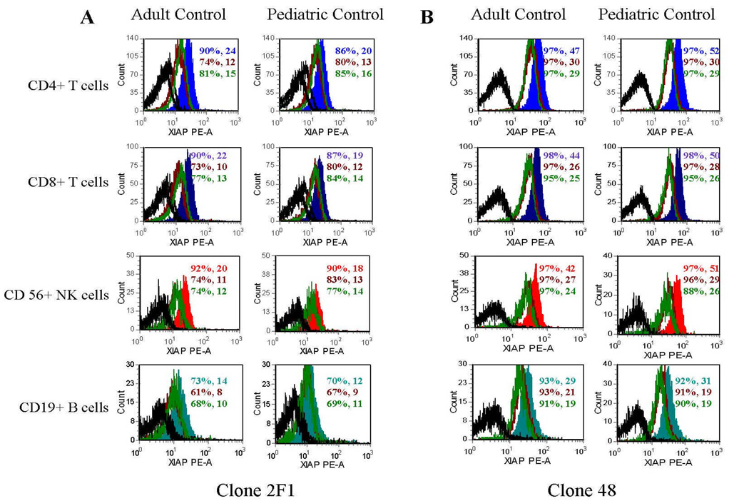
Methods: To develop a flow cytometric screening test for XIAP deficiency, we first used lymphoblastic cell lines generated from controls and patients with BIRC4 mutations to identify two commercially available antibodies specific for native intracellular XIAP. Next, we used these antibodies to study control whole blood leukocyte XIAP expression. We then studied XIAP expression in leukocytes from patients with XLP due to BIRC4 mutations, maternal carriers, and patients following HCT.

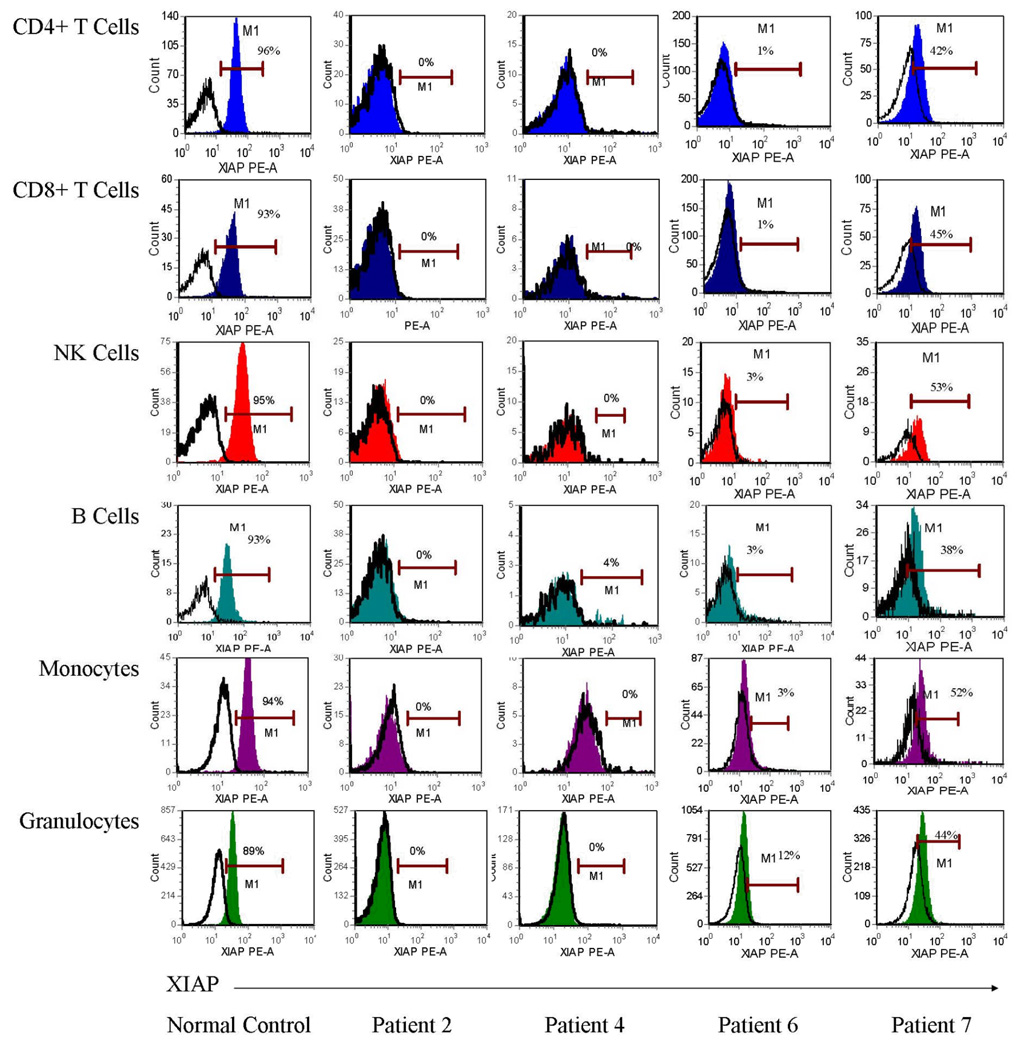
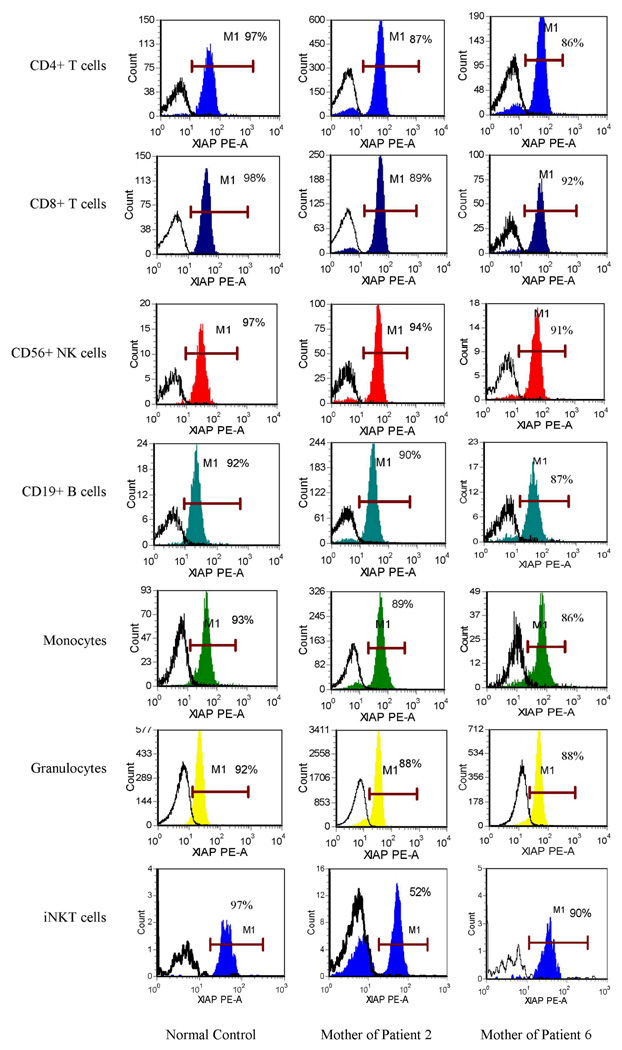
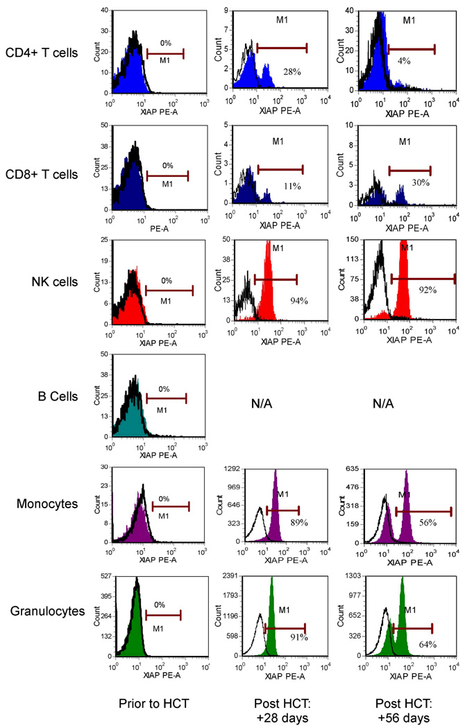
Results: XIAP was expressed by the majority of all whole blood nucleated cells in normal controls. In contrast, XIAP was absent or decreased in all lymphocyte subsets, monocytes and granulocytes from four unrelated patients with XLP due to BIRC4 mutations. Bimodal distribution of XIAP expression was evident in two maternal carriers, with significant skewing toward cells expressing normal XIAP. Bimodal distribution was also observed in a patient following HCT.

Conclusions: Flow cytometric analysis of intracellular XIAP provides a rapid screening test for XLP due to XIAP deficiency. It also allows carrier detection and can be used to monitor donor versus recipient reconstitution following HCT.


Figures

Figure 1
Figure 1
A schematic of the 6 exons of the BIRC4 gene and resultant protein, XIAP, with patient mutations included.
Figure 2
Figure 2
Analysis of XIAP expression within PBMCs and LCLs generated from patients with XIAP deficiency and normal controls. Figure A and B show western blot analysis of PBMC lysates of patients 1–4, probed with clone 28 anti-XIAP antibody (A) or clone 2F1 (B). Gels were loaded with 30 mcg of protein. Figures C and D depict LCL lysates from patients 1–5 using the clone 28 antibody. Figure C shows 30 mcg of protein loaded into the gel. Figure D shows the same samples, but with 5 mcg of protein loaded for control and patient 1, and 90mcg of protein loaded for patients 2–5. β-actin serves as a loading control. Figures E and F illustrate flow cytometric detection of XIAP in LCLs generated from patients 1–5, with 2 controls shown for comparison, using the clone 2F1 (E) or clone 48 (F) anti-XIAP antibodies. Debris was excluded from analysis by creating a viable LCL gate based on the appearance of forward and right angle light scatter. Filled histograms represent XIAP staining, while open histograms represent isotype control antibody. Percentages of cells positive for XIAP and the geometric mean channel fluorescence (gMCF) are given within the histogram boxes.
Figure 3
Figure 3
Flow cytometric detection of XIAP in peripheral whole blood granulocytes, monocytes, CD4+ T cells, CD8+ T cells, NK cells, and B cells, as described in the methods section, using the clone 48 antibody. Gating strategy is as shown. Filled histograms represent XIAP staining, while open histograms represent control antibody. Percentages of cells positive for XIAP and the geometric mean channel fluorescence (gMCF) are given within the histogram boxes.
Figure 4
Figure 4
Flow cytometric detection of XIAP in pediatric and adult peripheral blood CD4+ T cells, CD8+ T cells, NK cells, and B cells at 0, 12, and 36 hours after obtaining fresh blood samples. Colored filled histograms represent XIAP expression at 0 hours after blood samples were obtained, maroon unfilled histograms represent XIAP expression at 12 hours after blood samples obtained, and green unfilled histograms represent XIAP expression at 36 hours after blood samples were obtained. Black unfilled histograms represent isotype control antibody staining. 3A was obtained using the 2F1 antibody. 3B was obtained using the clone 48 antibody. Percentages of cells positive for XIAP and the gMCF are given within the histogram boxes.
Figure 5
Figure 5
Flow cytometric detection of XIAP in peripheral blood CD4+ T cells, CD8+ T cells, NK cells, B cells, monocytes and granulocytes from a representative normal control and XIAP deficient patients 2, 4, 6, and 7, using the clone 48 anti-XIAP antibody. Filled histograms represent XIAP staining, while open histograms represent control antibody.
Figure 6
Figure 6
Flow cytometric detection of XIAP in peripheral blood CD4+ T cells, CD8+ T cells, NK cells, B cells, monocytes, granulocytes and invariant NKT cells from a representative control and the mothers of patients 2 and 6 using the clone 48 anti-XIAP antibody. Filled histograms represent XIAP staining, while open histograms represent control antibody.
Figure 7
Figure 7
Flow cytometric detection of XIAP in peripheral blood CD4+ T cells, CD8+ T cells, NK cells, B cells, monocytes and granulocytes from XIAP deficient patient 2, prior to and at +28 and +56 days following allogeneic hematopoietic cell transplant (HCT) using the clone 48 anti-XIAP antibody. Filled histograms represent XIAP staining, while open histograms represent control antibody. N/A=not applicable, as there was no significant B-cell reconstitution at these time points.

References

    1. Rigaud S, Fondanèche MC, Lambert N, Pasquier B, Mateo V, Soulas P, et al. XIAP deficiency in humans causes an X-linked lymphoproliferative syndrome. Nature. 2006;444:110–114. - PubMed
    1. Tabata Y, Villanueva J, Lee SM, Zhang K, Kanegane H, Miyawaki T, et al. Rapid detection of intracellular SH2D1A protein in cytotoxic lymphocytes from patients with X-linked lymphoproliferative disease and their family members. Blood. 2005;105:3066–3071. - PubMed
    1. Kogawa K, Lee SM, Villanueva J, Marmer D, Sumegi J, Filipovich AH. Perforin expression in cytotoxic lymphocytes from patients with hemophagocytic lymphohistiocytosis and their family members. Blood. 2002;99:61–66. - PubMed
    1. Tosato G, Cohen JI. Generation of Epstein-Barr Virus (EBV)–Immortalized B Cell Lines. Curr Protoc Immunol. 2007 sup 76:Unit 7.22. - PubMed
    1. Duckett CS, Nava VE, Gedrich RW, Clem RJ, Van Dongen JL, Gilfillan MC, et al. A conserved family of cellular genes related to baculovirus iap gene and encoding apoptosis inhibitors. EMBO. 1996;15:2685–2694. - PMC - PubMed

Publication types

MeSH terms

Substances

LinkOut - more resources