. 2009 Mar 18;96(6):2194-203.
doi: 10.1016/j.bpj.2008.11.063.

Structure and membrane interactions of the antibiotic peptide dermadistinctin K by multidimensional solution and oriented 15N and 31P solid-state NMR spectroscopy

Affiliations

Structure and membrane interactions of the antibiotic peptide dermadistinctin K by multidimensional solution and oriented 15N and 31P solid-state NMR spectroscopy

Rodrigo M Verly et al. Biophys J. .

Abstract

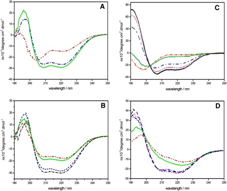
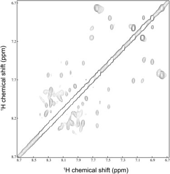
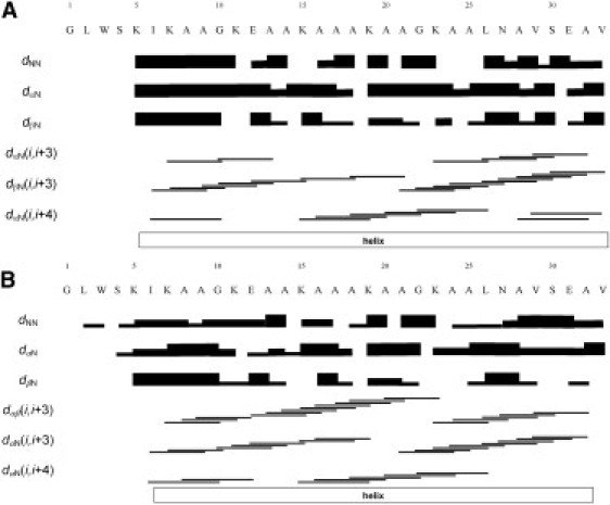
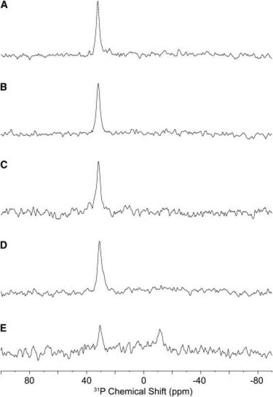
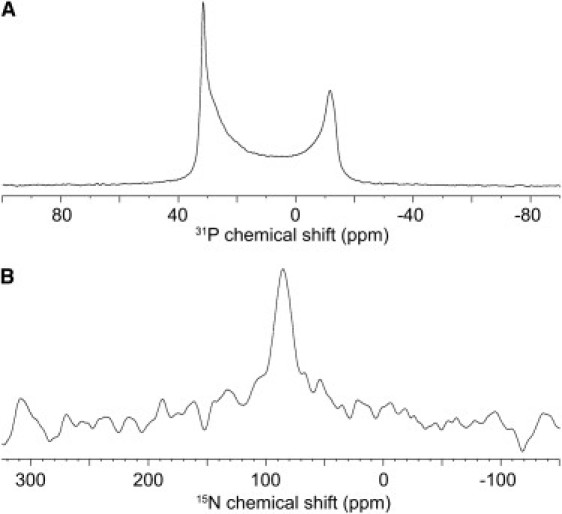
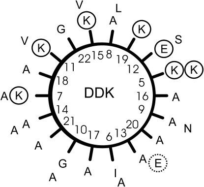
DD K, a peptide first isolated from the skin secretion of the Phyllomedusa distincta frog, has been prepared by solid-phase chemical peptide synthesis and its conformation was studied in trifluoroethanol/water as well as in the presence of sodium dodecyl sulfate and dodecylphosphocholine micelles or small unilamellar vesicles. Multidimensional solution NMR spectroscopy indicates an alpha-helical conformation in membrane environments starting at residue 7 and extending to the C-terminal carboxyamide. Furthermore, DD K has been labeled with (15)N at a single alanine position that is located within the helical core region of the sequence. When reconstituted into oriented phosphatidylcholine membranes the resulting (15)N solid-state NMR spectrum shows a well-defined helix alignment parallel to the membrane surface in excellent agreement with the amphipathic character of DD K. Proton-decoupled (31)P solid-state NMR spectroscopy indicates that the peptide creates a high level of disorder at the level of the phospholipid headgroup suggesting that DD K partitions into the bilayer where it severely disrupts membrane packing.


Figures

Figure 1
Figure 1
(A) CD spectra of 42 μM DD K in 100 mM NaCl, 20 mM phosphate buffer, pH 7 (dash, dot-dot), and after addition of 200 μM SDS (dash, dot) or 1 mM SDS (solid line). (B) CD spectra of 42 μM DD K in the presence of 200 μM DPC (dash, dot-dot), 400 μM DPC (solid line), 1 mM DPC (dash-dash), or 3 mM DPC (dash, dot). (C) CD spectra of 55 μM DD K in water (solid light line), 10% TFE (dash, dot-dot), 20% TFE (dash, dot), 30% TFE (dash, dot-dot-dot), and 60% TFE (solid black line). (D) CD spectra of 40 μM DD K in from (top to bottom) 10 μM POPG in the form of small unilamellar vesicles (dash, dot-dot), 20 μM POPG (solid line), 50 μM POPG (dash, dot), 100 μM POPG (dash-dash), and 200 μM POPG (dash, dot-dot).
Figure 2
Figure 2
NH-HN region of the NOESY spectrum of 1 mM DD K in a micellar solution of 400 mM DPCd38/H2O 5% D2O.
Figure 3
Figure 3
Graphical presentation of the NOEs observed for (A) 4 mM DD K in TFE/water 50:50 v/v, and (B) 1 mM DD K in 400 mM DPCd38/H2O and 5% D2O, pH 6. The dNN, dαN, and dβN are sequential NOEs, where strong and weak crosspeaks are indicated by thick and thin lines, respectively. The dαN (i, i + 3), dαβ(i, i + 3), and dαN (i, i + 4) medium-size connectivities link atoms of residues spaced as indicated by the horizontal lines. The outlines of the helical conformation are also indicated.
Figure 4
Figure 4
Solution NMR structures of DD K in (A) TFE/water 50:50 v/v, and in (B–D) a micellar solution of DPCd38 in water. (A and B) The 20 lowest energy structures. The hydrophobic residues are represented in blue, the hydrophilic residues in green. (C and D) The lowest energy structure in the presence of detergent viewed along the helix axis and from the side, respectively.
Figure 5
Figure 5
Proton-decoupled 31P solid-state NMR spectra of POPC bilayers in the presence of DD K as a function of peptide concentration: (A) in the absence of peptide; and in the presence of (B) 0.5 mol %; (C) 1 mol %; (D) 2 mol %; and (E) 3 mol % DD K. The bilayers are oriented with the membrane normal parallel to the magnetic field direction.
Figure 6
Figure 6
DD K labeled with 15N at its Ala-17 position and reconstituted at 1 mol % into oriented POPC bilayers. (A) Proton-decoupled 31P NMR and (B) proton-decoupled 15N solid-state NMR spectrum of the same sample.
Figure 7
Figure 7
Amphipathic helical wheel of DD K, residues 5–33. The inner circle shows amino acids 5–23, the outer residues 24–33. The charged side chains are circled. It remains possible that the polar C-terminus, including residue E31 is unstructured or distorted (hatched circle) in lipid bilayers thereby providing a more amphipathic structure to the polypeptide.

References

    1. Zasloff M. Antimicrobial peptides of multicellular organisms. Nature. 2002;415:389–395. - PubMed
    1. Boman H.G. Antibacterial peptides: basic facts and emerging concepts. J. Intern. Med. 2003;254:197–215. - PubMed
    1. Powers J.P., Hancock R.E. The relationship between peptide structure and antibacterial activity. Peptides. 2003;24:1681–1691. - PubMed
    1. Lehrer R.I. Primate defensins. Nat. Rev. Microbiol. 2004;2:727–738. - PubMed
    1. Garcia-Olmedo F., Molina A., Alamillo J.M., Rodriguez-Palenzuela P. Plant defense peptides. Biopolymers. 1999;47:479–491. - PubMed

Publication types

LinkOut - more resources