

Uniform action potential repolarization within the sarcolemma of in situ ventricular cardiomyocytes
- PMID: 19289075
- PMCID: PMC2907679
- DOI: 10.1016/j.bpj.2008.12.3896
Uniform action potential repolarization within the sarcolemma of in situ ventricular cardiomyocytes
Abstract
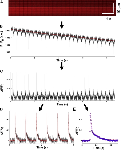
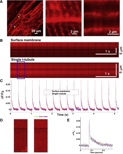
Previous studies have speculated, based on indirect evidence, that the action potential at the transverse (t)-tubules is longer than at the surface membrane in mammalian ventricular cardiomyocytes. To date, no technique has enabled recording of electrical activity selectively at the t-tubules to directly examine this hypothesis. We used confocal line-scan imaging in conjunction with the fast response voltage-sensitive dyes ANNINE-6 and ANNINE-6plus to resolve action potential-related changes in fractional dye fluorescence (DeltaF/F) at the t-tubule and surface membranes of in situ mouse ventricular cardiomyocytes. Peak DeltaF/F during action potential phase 0 depolarization averaged -21% for both dyes. The shape and time course of optical action potentials measured with the water-soluble ANNINE-6plus were indistinguishable from those of action potentials recorded with intracellular microelectrodes in the absence of the dye. In contrast, optical action potentials measured with the water-insoluble ANNINE-6 were significantly prolonged compared to the electrical recordings obtained from dye-free hearts, suggesting electrophysiological effects of ANNINE-6 and/or its solvents. With either dye, the kinetics of action potential-dependent changes in DeltaF/F during repolarization were found to be similar at the t-tubular and surface membranes. This study provides what to our knowledge are the first direct measurements of t-tubule electrical activity in ventricular cardiomyocytes, which support the concept that action potential duration is uniform throughout the sarcolemma of individual cells.
Figures


References
-
- Tidball J.G., Smith R., Shattock M.J., Bers D.M. Differences in action potential configuration in ventricular trabeculae correlate with differences in density of transverse tubule-sarcoplasmic reticulum couplings. J. Mol. Cell. Cardiol. 1988;20:539–546. - PubMed
-
- Blatter L.A., Niggli E. Confocal near-membrane detection of calcium in cardiac myocytes. Cell Calcium. 1998;23:269–279. - PubMed
-
- Swift F., Strømme T.A., Amundsen B., Sejersted O.M., Sjaastad I. Slow diffusion of K+ in the T tubules of rat cardiomyocytes. J. Appl. Physiol. 2006;101:1170–1176. - PubMed
Publication types
MeSH terms
Substances
Grants and funding
LinkOut - more resources
Full Text Sources
Other Literature Sources
Molecular Biology Databases
