Review
. 2009 Jun;1790(6):431-44.
doi: 10.1016/j.bbagen.2009.03.007. Epub 2009 Mar 14.

Regulation of integrin activity and signalling

Affiliations
Review

Regulation of integrin activity and signalling

Carl G Gahmberg et al. Biochim Biophys Acta. 2009 Jun.

Abstract

The ability of cells to attach to each other and to the extracellular matrix is of pivotal significance for the formation of functional organs and for the distribution of cells in the body. Several molecular families of proteins are involved in adhesion, and recent work has substantially improved our understanding of their structures and functions. Also, these molecules are now being targeted in the fight against disease. However, less is known about how their activity is regulated. It is apparent that among the different classes of adhesion molecules, the integrin family of adhesion receptors is unique in the sense that they constitute a large group of widely distributed receptors, they are unusually complex and most importantly their activities are strictly regulated from the inside of the cell. The activity regulation is achieved by a complex interplay of cytoskeletal proteins, protein kinases, phosphatases, small G proteins and adaptor proteins. Obviously, we are only in the beginning of our understanding of how the integrins function, but already now fascinating details have become apparent. Here, we describe recent progress in the field, concentrating mainly on mechanistical and structural studies of integrin regulation. Due to the large number of articles dealing with integrins, we focus on what we think are the most exciting and rewarding directions of contemporary research on cell adhesion and integrins.


Figures

Fig. 1
Fig. 1
The integrin superfamily. The integrins can be subdivided according to their β chains but note that some α chains can combine with several β chains. 24 different integrins are present in humans.
Fig. 2
Fig. 2
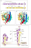
Structures of integrins and their extracellular domains. A, schematic drawing of the LFA-1 integrin. Note that the α I -domain is within the β-propeller, and the β I-like domain within the hybrid domain. The positions of the Mg++ (★) and Ca++ (★) binding sites are indicated; B, structure of the Mac-1 I-domain in the inactive state. The functionally important α1 (blue) and α7 (red) helices are shown. C, structure of the activated Mac-1 I-domain; note the magnesium ion (black dot) and the shifted position of the α7 helix. D, schematic structure of the external portion of αvβ3. In the crystal structure, the ligand binding site is turned towards the membrane (left); to the right is shown the stretched-out intermediate affinity form.
Fig. 3
Fig. 3
Valency and affinity modulations of integrins. By clustering of the integrins the avidity becomes high enough for functional adhesion (left). An intermediate affinity may be achieved by straightening out of the integrin, but high affinity needs opening of the binding site (right). It should be pointed out that it is not known for sure that the integrins need to straighten out in order to exhibit high affinity.
Fig. 4
Fig. 4
The cytoplasmic sequences of the human a and β integrin chains (β4 is not shown). The potential phosphorylation sites are marked in red. The established phosphorylation sites are numbered. The conserved membrane proximal sequences in the α chain are marked in blue. The functionally important NPXY(F) sequences in the β chain are marked in green and the threonine containing important phosphorylation sites in magenta.
Fig. 5
Fig. 5
Schematic structures of ICAMs. Similar Ig-like domains are colour coded. ICAM-1 and ICAM-3 are dimers, ICAM-2 and ICAM-4 monomers. ICAM-5 may exist as a dimer or tetramer.
Fig. 6
Fig. 6
Overlapping binding sites for cytoskeletal proteins in the β2 cytoplasmic segment. The functionally important threonine-758 residue is shown in bold. Phosphorylation of this residue enables binding of 14-3-3 proteins but inhibits filamin binding.
Fig. 7
Fig. 7
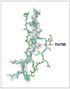
The crystal structure of the filamin domain 21 (green) binding region in complex with the β2 peptide. The important Thr-758 is shown. When this becomes phosphorylated hydrophobic interactions are disturbed and there is no space for the peptide in the filamin binding site and binding becomes impossible.
Fig. 8
Fig. 8
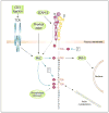
Phosphorylation of the β2 chain. Phosphorylation of β2 through the T cell receptor results in downstream events affecting integrin activation through avidity and affinity modulations.
Fig. 9
Fig. 9
A schematic and partially hypothetical view of LFA-1 activation. In the resting state (left) filamin is bound to the integrin and the ligand binding site is closed. Upon activation through the T cell receptor, talin may be cleaved by activated calpain, which results in binding of the talin head to the integrin cytoplasmic tails and separation of the integrin chains. Then phosphorylation of β2 (Thr-758) results in 14-3-3 binding, displacement of talin and activation of Rac1/Cdc42, which affect the actin cytoskeleton resulting in integrin clustering. The integrin is here shown in extended forms.

References

    1. Takeichi M. Cadherin cell adhesion receptors as a morphogenetic regulator. Science. 1991;251:1451–1455. - PubMed
    1. González-Amaro R, Sanchez-Madrid F. Cell adhesion molecules: selectins and integrins. Crit Rev Immunol. 1999;19:389–429. - PubMed
    1. Yona S, Lin HH, Siu WO, Gordon S, Stacey M. Adhesion-GPCRs: emerging roles for novel receptors. Trends Biochem Sci. 2008;33:491–500. - PubMed
    1. Hynes RO, Yamada KM. Fibronectins: multifunctional molecular glycoproteins. J Cell Biol. 1982;95:369–377. - PMC - PubMed
    1. Cunningham BA, Hemperly JJ, Murray BA, Prediger EA, Brackenbury R, Edelman GM. Neural cell adhesion molecule: structure, immunoglobulin-like domains, cell surface modulation, and alternative RNA splicing. Science. 1987;236:799–806. - PubMed

Publication types

LinkOut - more resources