. 2009 Jun;58(6):1302-11.
doi: 10.2337/db08-1113. Epub 2009 Mar 16.

Inhibition of Th17 cells regulates autoimmune diabetes in NOD mice

Affiliations

Inhibition of Th17 cells regulates autoimmune diabetes in NOD mice

Juliet A Emamaullee et al. Diabetes. 2009 Jun.

Abstract

Objective: The T helper 17 (Th17) population, a subset of CD4-positive T-cells that secrete interleukin (IL)-17, has been implicated in autoimmune diseases, including multiple sclerosis and lupus. Therapeutic agents that target the Th17 effector molecule IL-17 or directly inhibit the Th17 population (IL-25) have shown promise in animal models of autoimmunity. The role of Th17 cells in type 1 diabetes has been less clear. The effect of neutralizing anti-IL-17 and recombinant IL-25 on the development of diabetes in NOD mice, a model of spontaneous autoimmune diabetes, was investigated in this study.

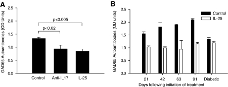
Research design and methods and results: Although treatment with either anti-IL-17 or IL-25 had no effect on diabetes development in young (<5 weeks) NOD mice, either intervention prevented diabetes when treatment was started at 10 weeks of age (P < 0.001). Insulitis scoring and immunofluorescence staining revealed that both anti-IL-17 and IL-25 significantly reduced peri-islet T-cell infiltrates. Both treatments also decreased GAD65 autoantibody levels. Analysis of pancreatic lymph nodes revealed that both treatments increased the frequency of regulatory T-cells. Further investigation demonstrated that IL-25 therapy was superior to anti-IL-17 during mature diabetes because it promoted a period of remission from new-onset diabetes in 90% of treated animals. Similarly, IL-25 delayed recurrent autoimmunity after syngeneic islet transplantation, whereas anti-IL-17 was of no benefit. GAD65-specific ELISpot and CD4-positive adoptive transfer studies showed that IL-25 treatment resulted in a T-cell-mediated dominant protective effect against autoimmunity.

Conclusions: These studies suggest that Th17 cells are involved in the pathogenesis of autoimmune diabetes. Further development of Th17-targeted therapeutic agents may be of benefit in this disease.


Figures

FIG. 1.
FIG. 1.
Inhibition of Th17 cells with either neutralizing anti–IL-17 or IL-25 has no effect when treatment is initiated during the initiation stage of autoimmune diabetes, but treatment significantly reduces the incidence of diabetes when it occurs during the effector stage. The impact of a neutralizing anti–IL-17 antibody as well as the Th17-inhibitory cytokine IL-25 was investigated during two phases of diabetes development in NOD mice. Treatment was initiated at either 5 weeks of age, which represents the initiation stage of autoimmunity, as characterized by mild insulitis, or at 10 weeks of age, when the effector phase of autoimmunity is occurring, and the majority of NODs will have invasive insulitis. Anti–IL-17 treatment (100 μg i.p. on days 0, 2, 4, 6, 8, and 10) did not alter the course of diabetes when it was initiated at 5 weeks of age (A), whereas treatment initiation at 10 weeks of age (B) resulted in a significant increase in the number of nondiabetic animals (P = 0.001 by log-rank). Similar results were obtained with recombinant IL-25, which had no effect when treatment was started at 5 weeks of age (C) but significantly increased diabetes-free survival at 10 weeks of age (D) (P = 0.001 by log-rank).
FIG. 2.
FIG. 2.
Both anti–IL-17 and IL-25 reduced the degree of insulitis in treated animals. A: Pancreata from anti–IL-17–treated, IL-25–treated, and control animals were harvested 1 month after the completion of treatment and stained with hematoxylin and eosin. The slides were blinded and scored by a pathologist according to the following scale (based on >50% of the islets in that section exhibiting the associated pattern): 0 (no infiltrate), 1 (mild peri-islet infiltrate), 2 (invasive insulitis with 25–50% islet destruction), and 3 (destructive insulitis with >50% islet destruction). Both anti–IL-17 (mean score 2.03 ± 0.33) and IL-25 (mean score 1.50 ± 0.33) treatment markedly reduced insulitis compared with controls (mean score 2.63 ± 0.26, P < 0.05 for anti–IL-17 vs. control and P < 0.02 for IL-25 vs. control by ANOVA). BD: Representative hematoxylin- and eosin-stained islets from each cohort (control [B], anti–IL-17 [C], and IL-25 [D]) are presented at ×200 magnification. (A high-quality digital representation of this figure is available in the online issue.)
FIG. 3.
FIG. 3.
Treatment with anti–IL-17 or IL-25 reduced the degree of peri-islet T-cell infiltration and was associated with an increase in the frequency of Foxp3-positive cells. Pancreata were collected from anti–IL-17–treated, IL-25–treated, and control animals 1 month after the completion of treatment. Tissue sections were stained for either CD4, CD8, Foxp3, or IL-17 (each in green) in combination with insulin (red) and nuclei (4′,6-diamidino-2-phenylindole in blue). Treatment with either anti–IL-17 or IL-25 reduced the frequency of both CD4- and CD8-positive T-cells and increased the number of Foxp3-positive Treg cells in the peri-islet infiltrate compared with controls. IL-17–positive staining was only visible in a small proportion of cells present within the insulitic lesion in control animals, and this frequency was further reduced after treatment with anti–IL-17 or IL-25. CD4, CD8, and Foxp3 staining was completed on cryosections, whereas IL-17 staining was completed on fixed sections, resulting in a difference in appearance on photography. Pancreata harvested from n = 6–8 normoglycemic animals from each treatment group were analyzed, and representative sections from each combination of staining are shown at ×200 magnification. (A high-quality digital representation of this figure is available in the online issue.)
FIG. 4.
FIG. 4.
Treatment with anti–IL-17 or IL-25 prevents GAD65 autoantibody formation. Serum samples were collected in anti–IL-17–treated, IL-25–treated, and control animals 1 month after the completion of treatment and analyzed for GAD65 autoantibodies using an ELISA. A: Both anti–IL-17 and IL-25 treatment regimens were associated with a significant reduction in the number of GAD65 IgG antibodies compared with control animals (P < 0.02 for anti–IL-17 vs. control and P < 0.005 for IL-25 vs. control by ANOVA). B: Animals treated with IL-25 were followed prospectively for GAD65 autoantibody development, and this treatment effectively prevented autoantibody formation >90 days after the initiation of treatment (P < 0.001 by ANOVA). A minimum of n = 4 animals were analyzed at each time point. OD, optical density.
FIG. 5.
FIG. 5.
Treatment with IL-25, but not anti–IL-17, can reverse new-onset diabetes and delay recurrent autoimmunity after syngeneic islet transplantation. Naïve spontaneously diabetic (blood glucose >18 mmol/l) NOD mice were randomly assigned to receive either anti–IL-17 (100 μg i.p. every other day), IL-25 (1 μg s.c. daily), or control (IgG for anti–IL-17, vehicle for IL-25). A: Treatment with anti–IL-17 did not reverse hyperglycemia after new-onset diabetes in NOD mice. B: Treatment with IL-25 resulted in a period of normoglycemia (mean 8.53 ± 2.77 days) in 9 of 10 animals, whereas none of the controls returned to normoglycemia (P < 0.0001 by ANOVA). One IL-25–treated animal experienced permanent remission beyond 100 days and after the discontinuation of IL-25 treatment at day 30 (data not shown). C: Although anti–IL-17 treatment did result in prolongation of syngeneic islet graft survival in 2 of 5 animals, no significant difference in recurrent autoimmunity was observed compared with IgG-treated controls. P = 0.346 by log-rank. D: Treatment with IL-25 delayed recurrent autoimmunity after syngeneic islet transplantation (mean survival time of 7.2 ± 0.2 days in IL-25–treated animals vs. 4.2 ± 0.8 days in vehicle-treated animals; P = 0.0013 by log-rank).
FIG. 6.
FIG. 6.
IL-25 treatment reduces the frequency of autoreactive Th2 cells and Th17 cells and leads to the formation of a CD4-positive splenocyte population that can prevent type 1 diabetes development in an adoptive transfer model. At 1 month after the completion of treatment, splenocytes were harvested from either anti–IL-17–treated, IL-25–treated, or control animals (n = 3–4 animals per treatment group; all normoglycemic) that had been previously treated beginning at 10 weeks of age. Purified splenocytes were analyzed using cytokine ELISpot assays or adoptively transferred into NOD-RAG−/− recipients. A: No difference in the frequency of GAD65-responsive, IFN-γ–secreting splenocytes was observed in either treatment group versus controls. B: IL-25 treatment resulted in a reduction in the number of GAD65-responsive, IL-4–secreting splenocytes compared with both anti–IL-17–treated animals (*P < 0.02 for IL-25 vs. anti–IL-17 and control by ANOVA), suggesting that IL-25 treatment can reduce the frequency of autoreactive Th2 cells. C: Whereas IL-25 treatment was associated with a reduction in the number of GAD65-responsive, IL-17–secreting splenocytes (# P < 0.05 vs. control by ANOVA), anti–IL-17 treatment significantly increased the frequency of this population (*P < 0.001 by ANOVA vs. control and IL-25). D: CD4-positive lymphocytes were further purified from these splenocyte preparations using magnetic beads. Naïve NOD-RAG−/− males received either 1 × 10⁷ splenocytes harvested from spontaneously diabetic NOD mice (diabetic splenocytes) combined with 2 × 10⁶ CD4-positive splenocytes isolated from mice previously treated with anti–IL-17, 1 × 10⁷ diabetic splenocytes combined with 2 × 10⁶ CD4-positive splenocytes from mice previously treated with IL-25, or 1 × 10⁷ diabetic splenocytes with no CD4 supplementation (control). Animals were monitored three times per week thereafter for diabetes onset. Supplementation with CD4-positive cells harvested from mice previously treated with IL-25 resulted in a significant dominant protective effect, preventing diabetes development in 75% of the animals in this group (P < 0.005 vs. anti–IL-17 and control by log-rank). Supplementation with CD4-positive cells from animals previously treated with anti–IL-17 had no effect, with 100% of the animals becoming diabetic within 60 days post-transfer, which was comparable to the diabetes incidence observed in the control group.
FIG. 7.
FIG. 7.
Treatment with anti–IL-17 or IL-25 increases the frequency of Treg cells within pancreatic lymph nodes. At 1 month after the completion of treatment, pancreatic draining lymph nodes or spleens were harvested from either anti–IL-17–treated, IL-25–treated, or control animals (n = 3–4 animals per treatment group), and lymphocytes were extracted. Purified lymphocytes were analyzed using flow cytometry and TGF-β1 ELISA to quantify the Treg cell activity present in each immune compartment. A: Representative flow cytometry panels from pancreatic lymph node cells harvested from each treatment group are shown. Each panel was first gated on lymphocytes and CD4-positive cells, and the dot plots shown compare CD25 staining (y-axis) versus FoxP3 staining (x-axis). B: The percentage of CD4-, CD25-, and FoxP3-positive Treg cells derived from spleen and pancreatic lymph nodes are shown. Treatment with either anti–IL-17 or IL-25 resulted in a significant increase in the proportion of Treg cells present in both the spleen and the pancreatic lymph node. # P < 0.05 vs. vehicle. C: After purification, 2 × 10⁵ cells from each animal in each treatment group were incubated with GAD65 for 48 h, and the supernatants were subsequently harvested for analysis using TGF-β1 ELISA. Treatment with anti–IL-17 or IL-25 resulted in a significant increase in TGF-β1 among pancreatic lymph node–derived lymphocytes compared with controls, whereas no difference was observed in splenic lymphocytes. *P < 0.001 vs. vehicle; # P < 0.005 vs. anti–IL-17.

References

    1. Suarez-Pinzon WL, Rabinovitch A: Approaches to type 1 diabetes prevention by intervention in cytokine immunoregulatory circuits. Int J Exp Diabetes Res 2001; 2: 3– 17 - PMC - PubMed
    1. Hedegaard CJ, Krakauer M, Bendtzen K, Lund H, Sellebjerg F, Nielsen CH: T helper cell type 1 (Th1), Th2 and Th17 responses to myelin basic protein and disease activity in multiple sclerosis. Immunology 2008; 125: 161– 169 - PMC - PubMed
    1. Mouzaki A, Deraos S, Chatzantoni K: Advances in the treatment of autoimmune diseases; cellular activity, type-1/type-2 cytokine secretion patterns and their modulation by therapeutic peptides. Curr Med Chem 2005; 12: 1537– 1550 - PubMed
    1. van Roon JA, Bijlsma JW, Lafeber FP: Suppression of inflammation and joint destruction in rheumatoid arthritis may require a concerted action of Th2 cytokines. Curr Opin Investig Drugs 2002; 3: 1011– 1016 - PubMed
    1. Bettelli E, Korn T, Kuchroo VK: Th17: the third member of the effector T cell trilogy. Curr Opin Immunol 2007; 19: 652– 657 - PMC - PubMed

Publication types

MeSH terms