. 2009 May 8;284(19):13045-56.
doi: 10.1074/jbc.M808352200. Epub 2009 Mar 16.

Generating an unfoldase from thioredoxin-like domains

Affiliations

Generating an unfoldase from thioredoxin-like domains

Michele L Forster et al. J Biol Chem. .

Abstract

Protein-disulfide isomerase (PDI), an endoplasmic reticulum (ER)-resident protein, is primarily known as a catalyst of oxidative protein folding but also has a protein unfolding activity. We showed previously that PDI unfolds the cholera toxin A1 (CTA1) polypeptide to facilitate the ER-to-cytosol retrotranslocation of the toxin during intoxication. We now provide insight into the mechanism of this unfoldase activity. PDI includes two redox-active (a and a') and two redox-inactive (b and b') thioredoxin-like domains, a linker (x), and a C-terminal domain (c) arranged as abb'xa'c. Using recombinant PDI fragments, we show that binding of CTA1 by the continuous PDIbb'xa' fragment is necessary and sufficient to trigger unfolding. The specific linear arrangement of bb'xa' and the type a domain (a' versus a) C-terminal to bb'x are additional determinants of activity. These data suggest a general mechanism for the unfoldase activity of PDI: the concurrent and specific binding of bb'xa' to particular regions along the CTA1 molecule triggers its unfolding. Furthermore, we show the bb' domains of PDI are indispensable to the unfolding reaction, whereas the function of its a' domain can be substituted partially by the a' domain from ERp57 (abb'xa'c) or ERp72 (ca degrees abb'xa'), PDI-like proteins that do not unfold CTA1 normally. However, the bb' domains of PDI were insufficient to convert full-length ERp57 into an unfoldase because the a domain of ERp57 inhibited toxin binding. Thus, we propose that generating an unfoldase from thioredoxin-like domains requires the bb'(x) domains of PDI followed by an a' domain but not preceded by an inhibitory a domain.


Figures
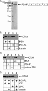
FIGURE 1.
FIGURE 1.
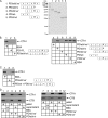
Recombinant PDI unfolds CTA1 efficiently. A, Coomassie stain of His-tagged full-length PDI (PDI-FL) purified from bacteria and subjected to SDS-PAGE. B, CTA was incubated with BSA or PDI in the presence of GSH at 30 °C followed by incubation with or without trypsin at 4 °C. Samples were resolved by reducing SDS-PAGE and analyzed by immunoblotting with an anti-CTA antibody that recognizes CTA1. C, as in B, except native PDI isolated from bovine liver was used, and all samples were treated with trypsin. D, as in B, except CTA was incubated with PDI at 4 or 30 °C before incubation with trypsin at 4 °C.
FIGURE 2.
FIGURE 2.
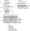
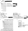
PDIbbxa′ is the minimum unit required to unfold CTA1. A, Coomassie stain of the indicated His-tagged PDI proteins purified from bacteria and subjected to SDS-PAGE. B, CTA was incubated with the indicated proteins in the presence of GSH followed by incubation with trypsin. Samples were analyzed by SDS-PAGE and immunoblotting with an anti-CTA antibody. C, CTA was incubated with PDIabbxa′ or an equal molar amount of PDIab and PDIbxa′ and analyzed as in B. D, CT holotoxin was noncovalently bound to GM1-coated beads, and the beads were incubated with the indicated proteins in the presence of GSH. After sedimenting the beads, the pellet and supernatant fractions were subjected to SDS-PAGE and immunoblot analysis with anti-CTA (top) or anti-CTB (bottom) antibodies.
FIGURE 3.
FIGURE 3.
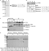
A PDI-toxin interaction is necessary but not sufficient to trigger unfolding. A, CTA was incubated with the indicated proteins in the presence of GSH to stimulate the unfolding reaction. PDI-toxin complexes were then isolated by co-immunoprecipitation with an antibody against the His tag on PDI and resolved by reducing SDS-PAGE. CTA1 was detected by immunoblot analysis using an anti-CTA antibody (top); ERp29 was detected by Coomassie stain (middle), and PDI was detected by immunoblotting with an anti-PDI antibody (bottom). B, as in A except PDI proteins were detected with Coomassie stain only (top). The intensities of the CTA1 bands were quantified with ImageJ (National Institutes of Health). Means ± S.D. (error bars) of four experiments are shown (bottom). C, CTA was incubated with the indicated proteins in the presence of GSH or GSSG. Immunoprecipitation and analysis were performed as in A.
FIGURE 4.
FIGURE 4.
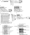
Linear arrangement and identity of domains are determinants of unfoldase activity. A, Coomassie stain of the indicated His-tagged proteins purified from bacteria and subjected to SDS-PAGE. B, CTA was incubated with the indicated proteins in the presence of GSH followed by incubation with trypsin. CTA1 was detected by SDS-PAGE followed by immunoblotting with an anti-CTA antibody. C, CTA was incubated with the indicated proteins in the presence of GSH to stimulate the unfolding reaction. PDI-toxin complexes were isolated by co-immunoprecipitation with an antibody against the His tag on PDI and resolved by reducing SDS-PAGE. CTA1 was detected by immunoblot analysis using an anti-CTA antibody (top), and PDI proteins were detected by Coomassie stain (bottom). D, Coomassie stain of the indicated proteins subjected to SDS-PAGE after incubation with the indicated amounts of trypsin.
FIGURE 5.
FIGURE 5.
Functional differences between the PDI and ERp57 thioredoxin-like domains. A, diagram of the domain architectures of full-length PDI and ERp57, designated FL for full-length. B, Coomassie stain of the indicated His-tagged proteins purified from bacteria and subjected to SDS-PAGE. C, CTA was incubated with the indicated proteins in the presence of GSH followed by incubation with trypsin. Samples were resolved by SDS-PAGE and immunoblotted with a CTA antibody. D, CT holotoxin was noncovalently bound to GM1-coated beads, and the beads were incubated with the indicated proteins in the presence of GSH. After sedimenting the beads, the pellet and supernatant fractions were subjected to SDS-PAGE and immunoblot analysis with anti-CTA (top) or anti-CTB (bottom) antibodies. E, Coomassie stain of the indicated proteins subjected to SDS-PAGE after incubation with the indicated amounts of trypsin. F, Coomassie stain of ERp57a-PDIbb′-ERp57xa′ purified from bacteria and subjected to SDS-PAGE. G, as in C. H, CTA was incubated with the indicated proteins in the presence of GSH. Recombinant proteins were then immunoprecipitated (IP) with an anti-His antibody and subjected to reducing SDS-PAGE. Co-precipitated CTA1 was detected by immunoblotting with an anti-CTA antibody (top), and the His-tagged proteins were detected by Coomassie stain (bottom). I, Coomassie stain of the indicated proteins subjected to SDS-PAGE after incubation with the indicated amounts of trypsin.
FIGURE 6.
FIGURE 6.
Functional differences between the PDI and ER72 thioredoxin-like domains. A, diagram of the domain architectures of full-length PDI and ERp72, designated FL for full-length. B, Coomassie stain of the indicated His-tagged proteins purified from bacteria and subjected to SDS-PAGE. C, CTA was incubated with the indicated proteins in the presence of GSH followed by incubation with trypsin. Samples were resolved by SDS-PAGE and immunoblotted with an anti-CTA antibody. D, Coomassie stain of the indicated proteins subjected to SDS-PAGE after incubation with the indicated amounts of trypsin. E, CTA was incubated with the indicated proteins in the presence of GSH to stimulate the unfolding reaction. Recombinant proteins were immunoprecipitated (IP) with an anti-His antibody and subjected to SDS-PAGE. Co-precipitated CTA1 was detected by immunoblotting with an anti-CTA antibody (top); PDI and the PDI-ERp72 hybrid were detected using an anti-PDI antibody (middle); and ERp29 was detected with an anti-ER29 antibody (bottom).

References

    1. Ferrari, D. M., and Soling, H. D. (1999) Biochem. J. 339 1-10 - PMC - PubMed
    1. Goldberger, R. F., Epstein, C. J., and Anfinsen, C. B. (1964) J. Biol. Chem. 239 1406-1410 - PubMed
    1. Gilbert, J., Ou, W., Silver, J., and Benjamin, T. (2006) J. Virol. 80 10868-10870 - PMC - PubMed
    1. Schelhaas, M., Malmstrom, J., Pelkmans, L., Haugstetter, J., Ellgaard, L., Grunewald, K., and Helenius, A. (2007) Cell 131 516-529 - PubMed
    1. Park, B., Lee, S., Kim, E., Cho, K., Riddell, S. R., Cho, S., and Ahn, K. (2006) Cell 127 369-382 - PubMed

Publication types

MeSH terms

LinkOut - more resources