. 2009 Dec;11(6):790-802.
doi: 10.1215/15228517-2009-015.

REV3L confers chemoresistance to cisplatin in human gliomas: the potential of its RNAi for synergistic therapy

Affiliations

REV3L confers chemoresistance to cisplatin in human gliomas: the potential of its RNAi for synergistic therapy

Huibo Wang et al. Neuro Oncol. 2009 Dec.

Abstract

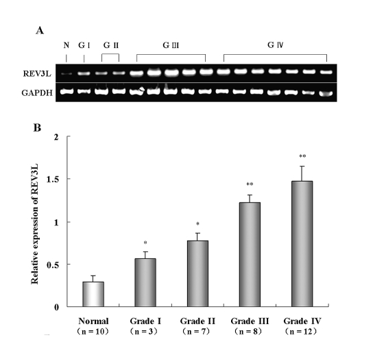
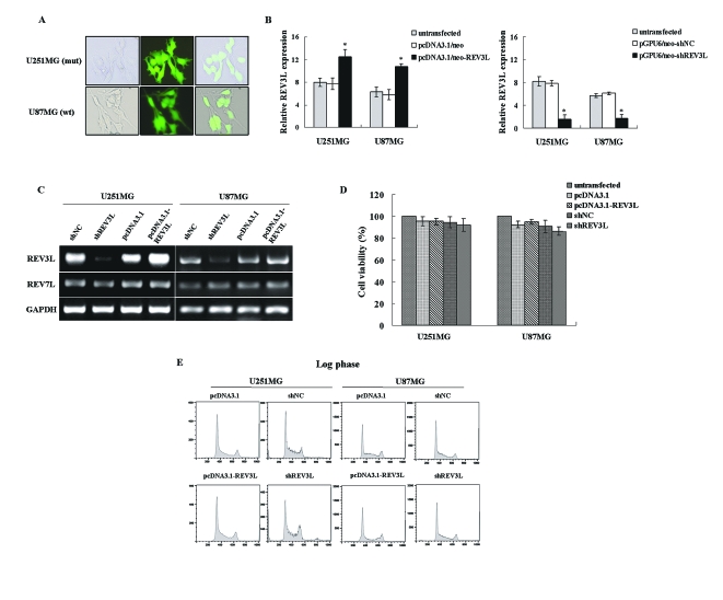
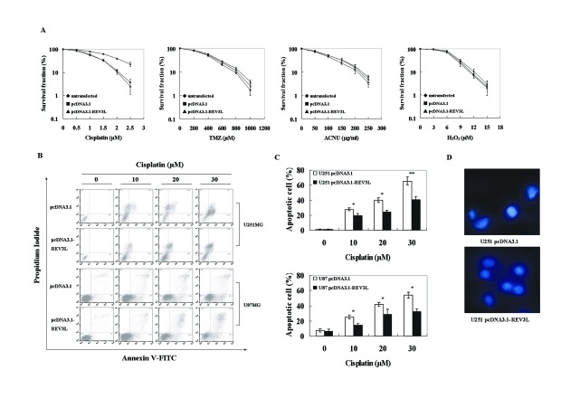
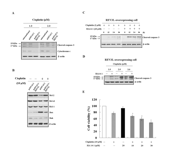
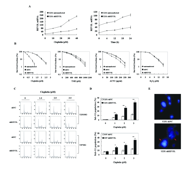
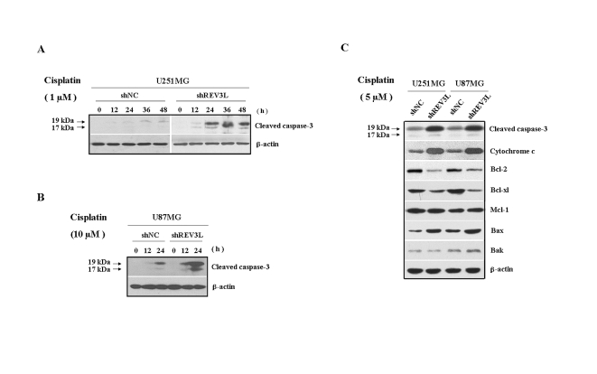
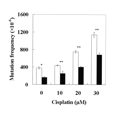
The REV3L gene, encoding the catalytic subunit of human polymerase zeta, plays a significant role in the cytotoxicity, mutagenicity, and chemoresistance of certain tumors. However, the role of REV3L in regulating the sensitivity of glioma cells to chemotherapy remains unknown. In this study, we investigated the expression of the REV3L gene in 10 normal brain specimens and 30 human glioma specimens and examined the value of REV3L as a potential modulator of cellular response to various DNA-damaging agents. Reverse transcriptase PCR/real-time PCR analysis revealed that REV3L was overexpressed in human gliomas compared with normal brain tissues. A glioma cell model with stable overexpression of REV3L was used to probe the role of REV3L in cisplatin treatment; upregulation of REV3L markedly attenuated cisplatin-induced apoptosis of the mitochondrial apoptotic pathway. We therefore assessed the REV3L-targeted treatment modality that combines suppression of REV3L expression using RNA interference (RNAi) with the cytotoxic effects of DNA-damaging agents. Downregulation of REV3L expression significantly enhanced the sensitivity of glioma cells to cisplatin, as evidenced by the increased apoptosis rate and marked alterations in the anti-apoptotic proteins B-cell lymphoma 2 (Bcl-2) and B-cell lymphoma-extra large (Bcl-xl) and proapoptotic Bcl-2-associated x protein (Bax) expression levels, and reduced mutation frequencies in surviving glioma cells. These results suggest that REV3L may potentially contribute to gliomagenesis and play a crucial role in regulating cellular response to the DNA cross-linking agent cisplatin. Our findings indicate that RNAi targeting REV3L combined with chemotherapy has synergistic therapeutic effects on glioma cells, which warrants further investigation as an effective novel therapeutic regimen for patients with this malignancy.


Figures

Fig. 1
Fig. 1
mRNA expression of REV3L in normal brain specimens and human glioma specimens. (A) Representative images of reverse transcriptase PCR analysis for REV3L gene expression in 15 specimens of human gliomas (G). REV3L gene expression was significantly elevated in malignant gliomas compared with normal brain tissue (N). (B) mRNA expression of REV3L measured by real-time PCR analysis. REV3L expression was significantly upregulated in malignant gliomas compared with normal brain tissues. Data are means of three independent experiments + SD. *p <0.01; **p <0.001.
Fig. 2
Fig. 2
Influence of upregulation or downregulation of REV3L on REV7L expression, cell proliferation, and cell cycle distribution in established glioma cells. (A) Representative images of stable transfectants of short hairpin interfering RNA for REV3L (shREV3L) U251 cells [U251MG (mt)] and shREV3L U87 cells [U87MG (wt)] in selection media under a fluorescence microscope. (B) Real-time PCR analysis for REV3L mRNA expression in U251 and U87 glioma cells. REV3L expression was significantly increased in the REV3L-overexpressing cells (left) and was significantly decreased in shREV3L cells (right) compared with control cells and untransfected cells. Data are means of three independent experiments ± SD. *p < 0.01. (C) Reverse transcriptase PCR analysis for REV3L and REV7L mRNA expression in short hairpin RNA-transfected negative control cells (shNC), shREV3L cells, vector control cells, and REV3L-overexpressing cells. Suppression or enhancement of REV3L expression in established cell lines did not influence REV7L mRNA expression. (D) The effect of REV3L expression on cell viability by diphenyltetrazolium bromide assay. Enhancement or suppression of REV3L expression did not have a significant effect on cell survival. Data are means of three independent experiments ± SD. (E) The effect of REV3L expression on cell cycle distribution by flow cytometry. Enhancement or suppression of REV3L expression did not affect cell cycle distribution.
Fig. 3
Fig. 3
REV3L overexpression rendered glioma cells resistant to DNA cross-linking agents. (A) Colony formation ability of the untransfected U251 cells, the vector control U251 cells, and the REV3L-overexpressing U251 cells after continuous treatment with cisplatin, temozolomide (TMZ), nimustine hydrochloride (ACNU), and H2O2. Overexpression of REV3L rendered glioma cells resistant to cisplatin but not to TMZ, ACNU, or H2O2. Data are means of three independent experiments ± SD. (B) Overexpression of REV3L inhibited apoptosis induced by cisplatin. Cells were treated with the indicated concentrations of cisplatin for 24 h, followed by staining with Annexin V–fluorescein isothiocyanate (FITC) and propidium iodide (PI) for the detection of dying (Annexin V+ PI) or dead (Annexin V+ PI+) cells. Apoptotic rates of the REV3L-overexpressing cells were significantly lower than those of the vector control cells exposed to similar conditions. (C) Apoptosis results of three independent experiments. Data are means of three independent experiments ± SD. *p <0.01; **p <0.001. (D) Morphological examination to detect apoptosis by 4′,6-diamidino-2-phenylindole dihydrochloride staining. Nuclear condensation and fragmentation of the REV3L-overexpressing U251 cells were not more prominent than vector control U251 cells after treatment with cisplatin (2 μmol/l) for 24 h.
Fig. 4
Fig. 4
REV3L overexpression conferred resistance to DNA cross-linking agents via inhibition of cisplatin-induced cell death of mitochondria-mediated apoptotic pathway. (A) The expression of cleaved caspase-3 and cytochrome c after exposure to two doses of cisplatin (1 and 2 μmol/l) for 48 h in untransfected U251 cells, vector control U251 cells, and REV3L-overexpressing U251 cells. Expression levels of cleaved caspase-3 and cytochrome c were lower in the REV3L-overexpressing U251 cells compared with the untransfected U251 cells and the vector control U251 cells. (B) Western blotting analysis of Bcl-2 family members in vector control U251 cells and REV3L-overexpressing U251 cells. Cells were cultured in the absence or presence of cisplatin (10 μmol/l) for 48 h. Expression levels of apoptosis-related effectors in treated cells did not significantly differ from those in untreated cells. The vector control cells that were treated with cisplatin (10 μmol/l) showed significant changes in the levels of Bcl-2 and Bax and a slight change in Bcl-xl. In contrast, the REV3L-overexpressing U251 cells treated with the same dose of cisplatin were indistinguishable from untreated cells, which suggests that no significant activation of these proteins occurred. Only a slight increase in Bax was observed in the REV3L-overexpressing U251 cells treated with cisplatin. (C) Effect of HA14-1 combined with cisplatin on the REV3L-overexpressing U251 cells. Cells were treated with cisplatin (2 μmol/l) in the absence or presence of HA14-1 (20 μmol/l) for the indicated periods. Expression levels of cleaved caspase-3 were significantly increased in REV3L-overexpressing U251 cells compared with cells treated with cisplatin only. (D) Effect of HA14-1 in combination with cisplatin on REV3L-overexpressing U251 cells. Cells were treated with HA14-1 (20 μmol/l) and three doses of cisplatin (1, 2, and 3 μmol/l) for 48 h. Expression levels of cleaved caspase-3 were significantly increased in REV3L-overexpressing U251 cells compared with levels in cells treated with cisplatin only. (E) Effect of HA14-1 treatment on cell viability after exposure to cisplatin by the diphenyltetrazolium bromide assay. Cells treated with HA14-1 alone were used as a toxicity control in the cell viability studies. HA14-1 treatment led to decreased cell viability in response to cisplatin. Data are means of three independent experiments ± SD. *p < 0.01.
Fig. 5
Fig. 5
REV3L knockdown can efficiently synergize the apoptosis response to treatment with cisplatin in glioma cells. (A) Real-time PCR analysis for REV3L mRNA level in the untransfected U251 cells and short hairpin interfering RNA for REV3L (shREV3L) U251 cells induced by various doses of cisplatin (10, 20, 30, and 40 μmol/l) at 24 h after a 1-h exposure (left), and time course change of REV3L mRNA level in both cell lines after exposure to 20 μmol/l cisplatin for 1 h (right). Data are means of three independent real-time PCR measurements ± SD. (B) Colony formation ability of the untransfected U251 cells, short hairpin RNA-transfected negative control (shNC) cells, and shREV3L cells after continuous treatment with cisplatin, temozolomide (TMZ), nimustine hydrochloride (ACNU), and H2O2. Suppression of REV3L expression conferred hypersensitivity to cisplatin but not to TMZ, ACNU, or H2O2. Data are means of three independent experiments ± SD. (C) Detection of apoptotic cells by flow cytometry. shNC U251, shREV3L U251, shNC U87, and shREV3L U87 cells were treated with three doses of cisplatin (1, 2, and 3 μmol/l) for 24 h. A sub-G1 peak was significantly higher in shREV3L cells than in shNC cells. (D) Sub-G1 peak results. Data are means of three independent experiments ± SD. *p < 0.01; **p < 0.001. (E) Morphological examination to detect apoptosis by 4′,6-diamidino-2-phenylindole dihydrochloride staining. Nuclear condensation and fragmentation of shREV3L cells were more prominent than shNC cells after treatment with cisplatin (2 μmol/l) for 24 h.
Fig. 6
Fig. 6
REV3L knockdown could efficiently synergize with cisplatin to induce glioma cell apoptosis via the mitochondria-mediated apop-totic pathway. (A) Time-dependent expression of cleaved caspase-3 after exposure to a single dose of cisplatin (1 μmol/l) in short hairpin RNA-transfected negative control (shNC) and short hairpin interfering RNA for REV3L (shREV3L) U251 cells. Expression levels of cleaved caspase-3 were significantly higher in shREV3L U251 cells compared with shNC U251 cells in response to the same dose of cisplatin. (B) Time-dependent expression of cleaved caspase-3 after exposure to a single dose of cisplatin (10 μmol/l) in shNC and shREV3L U87 cells. Expression levels of cleaved caspase-3 were significantly higher in shREV3L U87 cells compared with shNC U87 cells in response to the same dose of cisplatin. (C) The expression levels of cleaved caspase-3, cytochrome c, and Bcl-2 family members after exposure to a single dose of cisplatin (5 μmol/l) for 48 h in shNC and shREV3L cells. Expression levels of Bcl-2 and Bcl-xl were significantly lower and levels of cleaved caspase-3, cytochrome c, and Bax were significantly higher in shREV3L cells compared with shNC cells in response to the same dose of cisplatin. In contrast, the expression levels of Mcl-1 and Bak were indistinguishable from those of shNC cells.
Fig. 7
Fig. 7
REV3L knockdown reduced mutation frequency at the hypoxanthine guanine phosphoribosyl transferase locus (HPRT): frequency of cisplatin-induced generation of 6-thioguanine (6-TG) mutants after a 1-h exposure to one of three doses of cisplatin (10, 20, and 30 μmol/l) and 2 weeks of culture. Cisplatin-induced generation of 6-TG mutants was significantly reduced in short hairpin interfering REV3L RNA (shREV3L) cells compared with short hairpin RNA-transfected negative control (shNC) cells in response to cisplatin. Data are means of three independent experiments ± SD. *p < 0.01; **p < 0.001.

References

    1. Behin A, Hoang-Xuan K, Carpentier AF, Delattre JY. Primary brain tumours in adults. Lancet. 2003;361:323–331. - PubMed
    1. Furnari FB, Fenton T, Bachoo RM, et al. Malignant astrocytic glioma: genetics, biology, and paths to treatment. Genes Dev. 2007;21:2683–2710. - PubMed
    1. Barzon L, Zanusso M, Colombo F, Palu G. Clinical trials of gene therapy, virotherapy, and immunotherapy for malignant gliomas. Cancer Gene Ther. 2006;13:539–554. - PubMed
    1. DeAngelis LM. Brain tumors. N Engl J Med. 2001;344:114–123. - PubMed
    1. Okada H, Pollack IF. Cytokine gene therapy for malignant glioma. Expert Opin Biol Ther. 2004;4:1609–1620. - PubMed

Publication types

MeSH terms