. 2009 Jun;29(11):2982-96.
doi: 10.1128/MCB.00042-09. Epub 2009 Mar 16.

A mouse PRMT1 null allele defines an essential role for arginine methylation in genome maintenance and cell proliferation

Affiliations

A mouse PRMT1 null allele defines an essential role for arginine methylation in genome maintenance and cell proliferation

Zhenbao Yu et al. Mol Cell Biol. 2009 Jun.

Erratum in

Abstract

Protein arginine methyltransferase 1 (PRMT1) is the major enzyme that generates monomethylarginine and asymmetrical dimethylarginine. We report here a conditional null allele of PRMT1 in mice and that the loss of PRMT1 expression leads to embryonic lethality. Using the Cre/lox-conditional system, we show that the loss of PRMT1 in mouse embryonic fibroblasts (MEFs) leads to the loss of arginine methylation of substrates harboring a glycine-arginine rich motif, including Sam68 and MRE11. The loss of PRMT1 in MEFs leads to spontaneous DNA damage, cell cycle progression delay, checkpoint defects, aneuploidy, and polyploidy. We show using a 4-hydroxytamoxifen-inducible Cre that the loss of PRMT1 in MEFs leads to a higher incidence of chromosome losses, gains, structural rearrangements, and polyploidy, as documented by spectral karyotyping. Using PRMT1 small interfering RNA in U2OS cells, we further show that PRMT1-deficient cells are hypersensitive to the DNA damaging agent etoposide and exhibit a defect in the recruitment of the homologous recombination RAD51 recombinase to DNA damage foci. Taken together, these data show that PRMT1 is required for genome integrity and cell proliferation. Our findings also suggest that arginine methylation by PRMT1 is a key posttranslational modification in the DNA damage response pathway in proliferating mammalian cells.


Figures
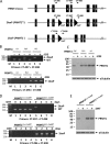
FIG. 1.
FIG. 1.
Generation of a PRMT1 conditional allele in mice and PRMT1-deficient MEFs. (A) Schematic representation of the wild-type (PRMT1 locus), floxed (2 loxP; PRMT1FL), and null (1 loxP; PRMT1) PRMT1 alleles. The exons are the black boxes, and the line represents introns not drawn to scale. The black triangles denote loxP sites, and the small arrows denote the primers (named CT#) used for PCR analysis. The expected size of the CT-497/498 DNA fragment for the wild-type allele is 245 bp, while the size of the DNA fragment for the 2loxP allele is 358 bp. The expected size of the CT-296/498 DNA fragment after CRE excision is 294 bp. (B) Primary MEFs deficient for PRMT1 were generated by infecting cells with a hygro-CRE retrovirus. Genomic DNA from hygromycin-selected cells was analyzed by PCR, and the DNA fragments were visualized on an ethidium bromide-stained agarose gel. M denotes molecular mass markers of the 1-kb ladder (Invitrogen, Inc.). (C) PRMT1 MEFs of the indicated genotype infected with hygro-Cre or not were lysed, and the total cellular proteins were analyzed by immunoblotting with anti-PRMT1 and anti-α-tubulin antibodies, as a loading control. The migration of PRMT1 and α-tubulin is shown on the right with arrows, and the migration of the molecular mass markers is shown on the left in kilodaltons. (D) PRMT1FL/− MEFs were immortalized and stably transfected with a plasmid encoding the estrogen receptor-CRE fusion protein (ER-CRE). The cells, termed PRMT1FL/−;CreERT MEFs or control PRMT1+/+;CreERT MEFs, were then treated with OHT for 0, 2, 4, and 6 days, and genomic DNA was isolated and analyzed as in panel B. (E) Total cellular proteins from mock-treated (−) or OHT-treated (+) PRMT1FL/−;CreERT and PRMT1+/+;CreERT MEFs were analyzed by immunoblotting as described for panel C.
FIG. 2.
FIG. 2.
Hypomethylation of cellular proteins in PRMT1-deficient MEFs. (A and B) PRMT1+/+ and PRMT1FL/− MEFs were left untreated (-CRE) or infected with the hygro-Cre retrovirus and selected with hygromycin (+CRE). Cell lysates were immunoblotted with anti-asymmetrical dimethylarginine antibodies ASYM24 (A) or ASYM25b (B) and anti-α-tubulin antibodies to control for equivalent loading. The migration of the molecular mass markers is shown on the left in kilodaltons. (C) PRMT1FL/− MEFs were left untreated (-CRE) or infected with hygro-Cre retroviruses (+CRE) and selected with hygromycin. Whole-cell lysates were immunoprecipitated with anti-Sam68 antibodies. The bound proteins were separated by SDS-polyacrylamide gel electrophoresis and immunoblotted with anti-Sam68 (left panel) and ASYM24 (right panel) antibodies as indicated. The migration of Sam68 and the heavy chain of IgG is shown. (D) PRMT1FL/−;CreERT MEFs were treated with (+OHT) or left untreated (-OHT) for 6 days. Whole-cell lysates were immunoprecipitated with anti-MRE11 antibodies, and the bound proteins were separated by SDS-polyacrylamide gel electrophoresis and immunoblotted with anti-MRE11 and ASYM25b antibodies as indicated. The migration of MRE11 is shown. (E) PRMT1FL/− MEFs were left untreated (-CRE) or infected with hygro-Cre retroviruses (+CRE) and selected with hygromycin. Whole-cell lysates were immunoblotted with the indicated PRMT antibodies or the anti-tubulin antibody to visualize equivalent loading.
FIG. 3.
FIG. 3.
PRMT1 is essential for the viability of MEFs. (A) The indicated primary PRMT1 MEFs (P3) were plated at cell density of 5 × 10⁵ cells per 10-cm tissue culture dish. Every two or 3 days, the cells were treated with trypsin and counted with a Beckman-Coulter counter, and a quarter of the cells were replated for the next counting (n = 4). (B) PRMT1FL/− MEFs were left uninfected (no infection) or infected with retroviruses encoding GFP (GFP) and a GFP-CRE (GFP-CRE) fusion protein, respectively. The GFP-positive cells were identified and counted by flow cytometry at 5 and 12 days postinfection. The y axis denotes the cell number, and the x axis denotes the GFP fluorescence. (C) The quantification of panel B expressed as a percentage of GFP-positive cells normalized to 100% with day 5. (D) Four hundred PRMT1FL/− (-ER-CRE) and PRMT1FL/−;CreERT (+ER-CRE) MEFs were seeded on 10-cm tissue culture dishes and maintained in regular medium (Dulbecco modified Eagle medium:10% serum) containing different combinations of blasticidin and OHT as indicated. Twelve days later, the cells were fixed, and cell colonies were stained with crystal violet.
FIG. 4.
FIG. 4.
PRMT1-deficient MEFs exhibit >4N DNA content and accumulation of cells at G2/M phase. (A) PRMT1FL/−;CreERT MEFs were treated with OHT for the indicated days and the cells were stained with PI and analyzed by cell sorting. The percentage of cells containing >4N DNA content is indicated. The experiments were performed three times, and a representative profile is shown. (B) Cell extracts from OHT-treated PRMT1FL/−;CreERT MEFs were immunoblotted with anti-PRMT1 and antitubulin antibodies to confirm the loss of PRMT1. (C) PRMT1+/+;CreERT MEFs were treated with OHT for the indicated days, and the cells were stained with PI and analyzed by cell sorting. The percentage of cells containing >4N DNA content is indicated.
FIG. 5.
FIG. 5.
PRMT1-deficient MEFs exhibit cell cycle delays. (A) PRMT1FL/−;CreERT MEFs were left untreated (-OHT) or treated with OHT (OHT 6 and 10 days) to generate PRMT1-deficient MEFs. The cells growing in log phase were incubated with 10 μM BrdU for 45 min, washed twice with complete medium, and then cultured in the absence of BrdU for the indicated times. The experiments were performed twice, and a representative profile is shown. (B) Histogram representation of BrdU-positive cells. The cells from the boxed area of panel A were represented as a histogram. The 2N and 4N DNA is indicated.
FIG. 6.
FIG. 6.
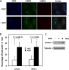
PRMT1-deficient MEFs exhibit spontaneous DNA damage. (A) PRMT1FL/−;CreERT MEFs were left untreated (-OHT) or treated with OHT for 6 days (+OHT). The cells were then visualized by indirect immunofluorescence microscopy using anti-γH2AX and anti-53BP1 antibodies. DAPI staining was used to stain the nucleus. (B) The percentage of cells with >5 foci was counted from three independent experiments. The statistical significance was assessed using the Student t test. (C) PRMT1FL/−;CreERT MEFs treated for 0, 4, and 8 days with OHT were lysed and analyzed by immunoblotting with anti-γH2AX and antitubulin antibodies.
FIG. 7.
FIG. 7.
Loss of PRMT1 in MEFs leads to genomic instability by SKY analysis. SKY was performed on the PRMT1FL/−;CreERT MEF cells untreated (-OHT) or treated with OHT for 6 days (+OHT). SKY was carried out as described in Materials and Methods. A summary of all chromosomal abnormalities for each cell line is given in Table 1. Two representative metaphase spreads of the +OHT cell line analyzed by SKY are shown. (A) Spectral karyotype of a metaphase with 142 chromosomes, represented in classification pseudocolors. (B) Spectral karyotype of a metaphase with 71 chromosomes. The karyotype is classified with the inverted-DAPI image (left) and the classification pseudocolors (right). Arrows indicate the chromosomal abnormalities that are found only in the +OHT cell line, including unique chromosome deletions and translocations, chromosome breaks, and dicentric chromosomes. The inverted-DAPI image of this metaphase is shown in Fig. S2 in the supplemental material.
FIG. 8.
FIG. 8.
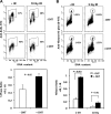
PRMT1 is required for both the G2/M and the G1/S checkpoint activations in response to IR-induced DNA damage. (A) Duplicate cultures of PRMT1FL/−;CreERT MEFs were left untreated (-OHT) or treated with OHT (+OHT). One set was treated with 10 Gy of IR (10 Gy IR), and the other set was left untreated (-IR). At 20 h after treatment, the cells were incubated with 10 μM BrdU for 1 h and stained with PI and an FITC-conjugated anti-BrdU antibody. The cells were analyzed by flow cytometry. A typical fluorescence-activated cell sorting profile is shown; the experiments were performed twice in duplicate, and the average is shown. The S-phase ratio was obtained by dividing the percentage of gated BrdU-positive cells treated with 10 Gy of IR by the percentage of gated BrdU-positive cells without IR treatment. Statistical significance was assessed by using Student t test. (B) Duplicate cultures of PRMT1FL/−;CreERT MEFs were left untreated (-OHT) or treated with OHT (+OHT). One set was treated with 2 Gy of IR (2 Gy IR), and the other set was left untreated (-IR). At 1.5 h after treatment, the cells were fixed and stained with PI and anti-pSer10-histone H3 antibody to identify the cells in mitosis. The percentage of pSer10-histone H3-positive cells (circled area) was determined by flow cytometry. The experiments were performed twice in duplicate. A typical result showing anti-pSer10-histone H3 antibody staining and the M-phase ratio, derived by dividing the percentage gated pSer10-histone H3-positive cells treated with 2 or 10 Gy of IR by the average percentage of gated pSer10-histone H3-positive cells without IR treatment. Statistical significance was assessed by using the Student t test.
FIG. 9.
FIG. 9.
U2OS cells with downregulated PRMT1 expression are hypersensitive to etoposide treatment. (A) U2OS cells were transfected with siPRMT1 and control siGFP, and the loss of PRMT1 expression was verified by immunoblotting. (B) U2OS cells were plated at 3 × 10⁵ cells/10-cm tissue culture dish and transfected with either siGFP or siPRMT1. The cell growth was monitored by counting with a Beckman-Coulter counter (n = 4). (C) Two days after transfection, the cells were treated with trypsin and 400 cells were replated on 10-cm tissue culture dishes. The cells were treated next day with etoposide with the desired dosage and time period as indicated. The cells were then washed twice with PBS and grown in regular medium. The visible colonies containing >20 cells were counted at 10, 12, and 14 days after treatment, respectively, and normalized to 100% without the treatment. Ten fields were randomly selected, and the total number of the colonies in the 10 fields was calculated and expressed as a percentage.
FIG. 10.
FIG. 10.
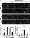
Downregulation of PRMT1 expression in U2OS cells leads to a reduced recruitment of RAD51 to DNA damage foci after IR treatment. U2OS cells were transfected with control siGFP and siPRMT1 and, 96 h after transfection, the cells were left untreated or treated with 10 Gy of IR. After various hours of recovery, the cells were visualized by indirect immunofluorescence with anti-53BP1 and anti-RAD51 antibodies. (A) Representative images. (B) Cells with >5 foci were counted and are expressed as a percentage. The graphs show the average and standard error of the mean from two independent experiments performed in duplicates where >10 different fields were analyzed. In total, more than 200 cells were counted for each sample in each experiment. Statistical significance was assessed by using the Student t test.

References

    1. Adams, M. M., B. Wang, Z. Xia, J. C. Morales, X. Lu, L. A. Donehower, D. A. Bochar, S. J. Elledge, and P. B. Carpenter. 2005. 53BP1 oligomerization is independent of its methylation by PRMT1. Cell Cycle 41854-1861. - PubMed
    1. Adelman, C. A., S. De, and J. H. Petrini. 2009. Rad50 is dispensable for the maintenance and viability of post-mitotic tissues. Mol. Cell. Biol. 29483-492. - PMC - PubMed
    1. An, W., J. Kim, and R. G. Roeder. 2004. Ordered cooperative functions of PRMT1, p300, and CARM1 in transcriptional activation by p53. Cell 117735-748. - PubMed
    1. Bachand, F. 2007. Protein arginine methyltransferases: from unicellular eukaryotes to humans. Eukaryot. Cell 6889-898. - PMC - PubMed
    1. Baumann, P., and S. West. 1998. Role of the human RAD51 protein in homologous recombination and double-stranded-break repair. Trends Biochem. Sci. 23247-251. - PubMed

Publication types

MeSH terms

LinkOut - more resources