. 2009 May;29(10):2748-61.
doi: 10.1128/MCB.01391-08. Epub 2009 Mar 16.

Trophoblast stem cell maintenance by fibroblast growth factor 4 requires MEKK4 activation of Jun N-terminal kinase

Affiliations

Trophoblast stem cell maintenance by fibroblast growth factor 4 requires MEKK4 activation of Jun N-terminal kinase

Amy N Abell et al. Mol Cell Biol. 2009 May.

Abstract

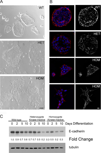
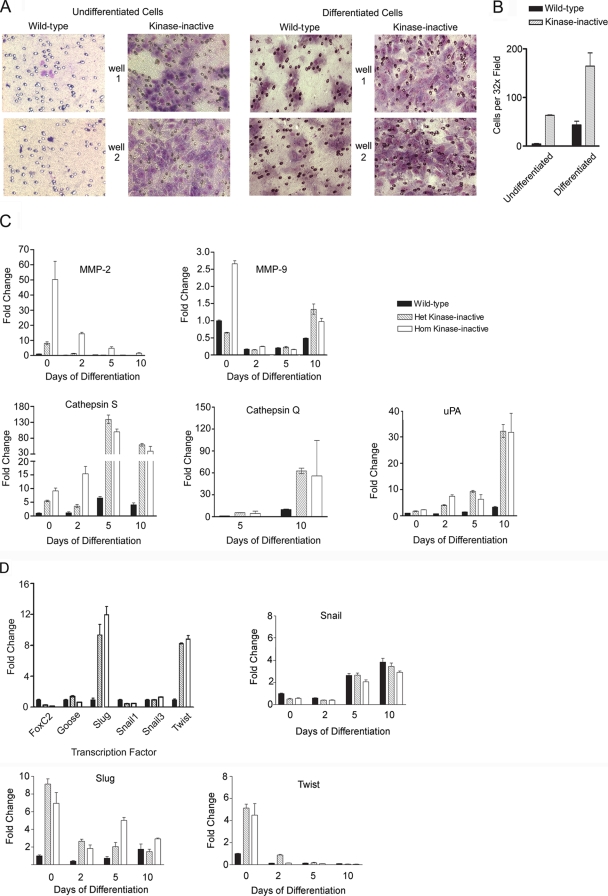
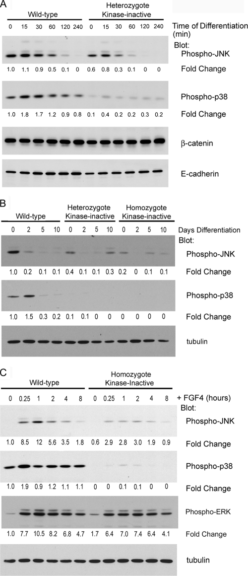
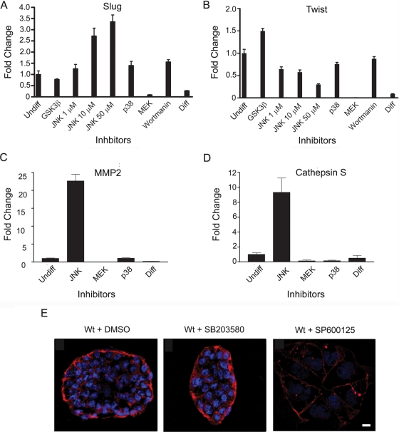
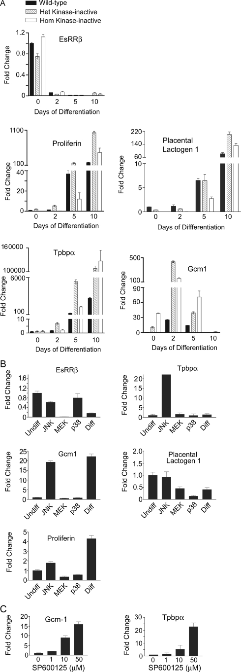
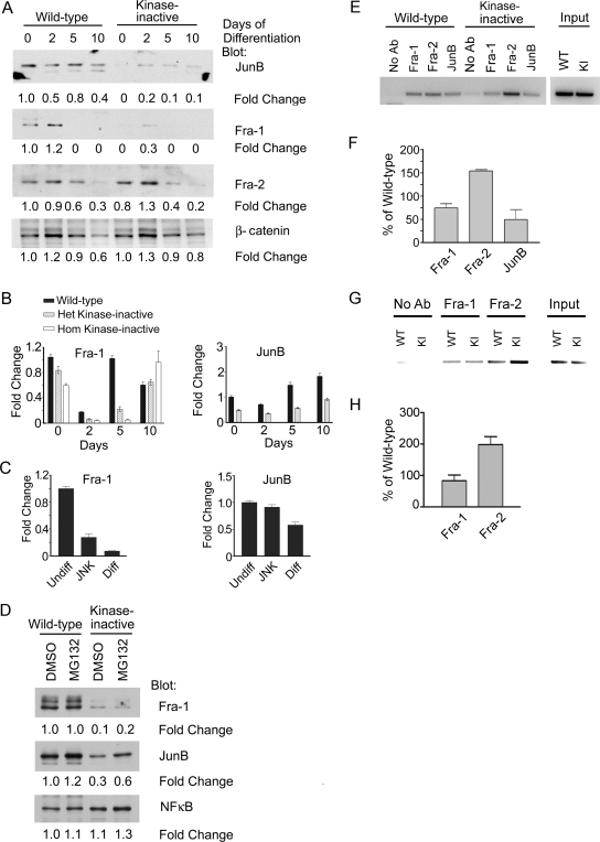
Trophoblast differentiation during placentation involves an epithelial-mesenchymal transition (EMT) with loss of E-cadherin and gain of trophoblast invasiveness. Mice harboring a point mutation that renders inactive the mitogen-activated protein kinase kinase kinase MEKK4 exhibit dysregulated placental development with increased trophoblast invasion. Isolated MEKK4 kinase-inactive trophoblast stem (TS) cells cultured under undifferentiating, self-renewing conditions in the presence of fibroblast growth factor 4 (FGF4) display increased expression of Slug, Twist, and matrix metalloproteinase 2 (MMP2), loss of E-cadherin, and hyperinvasion of extracellular matrix, each a hallmark of EMT. MEKK4 kinase-inactive TS cells show a preferential differentiation to Tpbp alpha- and Gcm1-positive trophoblasts, which are indicative of spongiotrophoblast and syncytiotrophoblast differentiation, respectively. FGF4-stimulated Jun N-terminal kinase (JNK) and p38 activity is markedly reduced in MEKK4 kinase-inactive TS cells. Chemical inhibition of JNK in wild-type TS cells induced a similar EMT response as loss of MEKK4 kinase activity, including inhibition of E-cadherin expression and increased expression of Slug, MMP2, Tpbp alpha, and Gcm1. Chromatin immunoprecipitation analyses revealed changes in AP-1 composition with increased Fra-2 and decreased Fra-1 and JunB binding to the regulatory regions of Gcm1 and MMP2 genes in MEKK4 kinase-inactive TS cells. Our results define MEKK4 as a signaling hub for FGF4 activation of JNK that is required for maintenance of TS cells in an undifferentiated state.


Figures

FIG. 1.
FIG. 1.
MEKK4 is expressed strongly in the trophoblast lineages of the developing placenta. MEKK4 was visualized in cryosections with anti-MEKK4 immunostaining (red) (A, C, D, H, and I) and by in situ hybridization with MEKK4 antisense probe (purple) (B, E, and F) or control MEKK4 sense probe (G). Nuclei were counterstained with DAPI (blue). (A) Anti-MEKK4 immunostaining shows that MEKK4 is expressed not only in the developing embryo but also in the invading primary giant cells lining the implantation site and the ectoplacental cone. (B) MEKK4 expression by in situ hybridization shows an identical expression pattern of MEKK4 message compared to the MEKK4 protein shown in panel A. (C and D) Strong MEKK4 expression in E8.5 chorion visualized by anti-MEKK4 immunostaining. The box indicates the area shown in the inset in panel D. (E) Strong expression of MEKK4 message in E8.5 chorion by in situ hybridization with antisense MEKK4 probe. (F) In situ hybridization with antisense MEKK4 probe in an E9.5 cryosection. (G) Control in situ hybridization with sense MEKK4 probe in an E9.5 cryosection. (H and I) Anti-MEKK4 immunostaining of E9.5 placenta (red). A white box indicates the area shown in the inset in panel I. For panels A to I, closed arrows indicate trophoblast giant cells, open arrows indicate spongiotrophoblasts, closed arrowheads indicate undifferentiated cuboidal trophoblasts, and open arrowheads indicate chorion. (J to O) Immunofluorescence microscopy of E9.5 placental cryosections stained with an anti-MEKK4 antibody (red) and counterstained with the nuclear stain DAPI (blue). (J to L) MEKK4 does not colocalize with PECAM-positive endothelial cells. Sections were also costained with anti-PECAM antibody (green). (M to O) MEKK4 colocalizes with cytokeratin (CK). Sections were also costained with anti-CK antibody. White boxes in panel J indicate the area that is enlarged in the insets in panels K and L. White boxes in panel M indicate areas enlarged in the insets in panels N and O. Filled arrowheads indicate MEKK4 in undifferentiated cuboidal trophoblasts. For panels J to O, filled arrows indicate PECAM-positive endothelial cells. Open arrows indicate sites with CK staining (green) having colocalization with MEKK4 (red). Bar, 100 μm.
FIG. 2.
FIG. 2.
Altered morphology of kinase-inactive (K1361R) placentas with dysregulated expression of Tpbpα. Placental sections from littermate E9.5 embryos with wild-type (MEKK4WT; left), heterozygous (MEKK4WT/K1361R; center), or homozygous kinase-inactive (MEKK4K1361R; right) genotypes. (A) Hematoxylin and eosin (H&E) staining of E9.5 placentas. White arrowheads indicate the location of the placenta. (B) In situ hybridization with antisense probe to the spongiotrophoblast marker Tpbpα, showing dysregulated expression of Tpbpα. Open black arrows indicate sites of Tpbpα expression. (C) In situ hybridization with antisense probe to the primary giant cell marker placental lactogen I (PlI). Bar, 0.1 mm. MEKK4 is expressed strongly in trophoblast stem cells and expression is reduced with differentiation. (D) MEKK4 is expressed more strongly in stem cells than in fibroblasts. Thirty micrograms of TS cell or MEF lysate was probed with anti-MEKK4 or anti-ΜΕΚΚ3 antibodies. (E) MEKK4 expression is reduced by TS cell differentiation. TS cells were differentiated by removal of FGF4 and fibroblast-conditioned medium for the indicated times and probed with anti-MEKK4 or anti-α-tubulin antibodies.
FIG. 3.
FIG. 3.
Reduced E-cadherin expression in TS cells homozygous for MEKK4K1361R. (A) Phase microscopy of wild-type and mutant TS cells cultured under undifferentiating conditions in the presence of FGF4 and conditioned medium is shown. Wild-type TS cells colonies exist as tight epithelial sheets. Heterozygote MEKK4WT/K1361R TS cell colonies are more irregularly shaped and slightly differentiated. Homozygote MEKK4K1361R TS cell colonies appear differentiated with irregularly shaped, polarized cells. Bar, 100 μm. (B) E-cadherin expression in wild-type and kinase-inactive TS cells cultured under undifferentiating conditions. Cells were immunostained with anti-E-cadherin antibodies (red) and nuclei were counterstained with DAPI (blue). Strong peripheral E-cadherin staining was observed in wild-type TS cells. Peripheral E-cadherin staining was slightly decreased in heterozygote kinase-inactive TS cells and further decreased in homozygote kinase-inactive TS cells. (C) Western blotting of whole-cell extracts from wild-type and kinase-inactive TS cells. Blots were probed with anti-E-cadherin and antitubulin antibodies. Changes were expressed relative to undifferentiated wild-type TS cells. WT, wild-type cells; HET, heterozygote kinase-inactive cells; HOM, homozygote kinase-inactive cells. Bar, 10 μm. Data shown are representative results from three independent experiments.
FIG. 4.
FIG. 4.
Increased invasiveness of MEKK4 kinase-inactive TS cells. (A) Invasion of undifferentiated or differentiated wild-type and heterozygote kinase-inactive TS cells through growth factor-reduced Matrigel. Chambers were Wright-stained and 10× images from two transwell chambers are shown for each condition. (B) Quantitation of the TS cell invasion shown in panel A. Data shown are the means ± ranges of two independent experiments performed in duplicate. Five 32× fields were counted for each transwell chamber. (C) Increased expression of proteases in kinase-inactive TS cells. Message levels were measured by quantitative RT-PCR of TS cells differentiated for the indicated number of days. Data are the means ± ranges of three independent experiments performed in duplicate and normalized to undifferentiated wild-type levels. (D) Increased expression of EMT-inducing transcription factors Slug and Twist in MEKK4 kinase-inactive TS cells. TS cells were cultured under undifferentiating conditions in the presence of FGF4 and conditioned medium. Message levels were measured by quantitative RT-PCR. Data normalized to wild-type undifferentiated TS cell levels are the means ± ranges of two independent experiments performed in duplicate. Het, heterozygous; Hom, homozygous.
FIG. 5.
FIG. 5.
FGF4 stimulation of JNK phosphorylation is reduced in kinase-inactive TS cells. Western blot analysis of wild-type, heterozygote, and homozygote kinase-inactive TS cell extracts is shown. Blots were probed with antibodies against phospho-JNK, phospho-p38, α-tubulin, β-catenin, and E-cadherin. Changes are relative to wild-type TS cells (time zero). (A) Removal of FGF4 from TS cells results in a loss of JNK phosphorylation. FGF4 and conditioned medium was removed for the indicated times. Phospho-JNK levels decreased after an hour of differentiation. Phospho-JNK and phospho-p38 levels were significantly reduced in kinase-inactive TS cells. (B) Differentiation results in a loss of phospho-JNK protein and a slight enhancement in phospho-p38 protein levels. Kinase-inactive TS cells exhibited a 60 to 80% reduction in phospho-JNK levels and a nearly complete loss of phospho-p38. (C) FGF4-stimulated JNK phosphorylation is diminished in kinase-inactive TS cells. TS cell were cultured for 24 h in the absence of FGF4 in TS medium containing TGF-β. FGF4 (10 ng/ml) was added for the indicated times. Data shown are representative results from three independent experiments.
FIG. 6.
FIG. 6.
Chemical inhibition of JNK induces the expression of Slug, MMP2, and cathepsin S and a loss of E-cadherin expression. (A, B, C, and D) Wild-type TS cells were cultured under undifferentiating conditions in the presence of FGF4 and conditioned medium and treated for 2 days with either DMSO, 30 μM GSK3β inhibitor, JNK inhibitor, 10 μM p38 inhibitor, 20 μM MEK inhibitor, 0.1 μM wortmannin, or differentiated (Diff) for 2 days in the absence of FGF4 and conditioned medium. Message levels were measured by quantitative RT-PCR. Data were normalized to wild-type undifferentiated DMSO-treated TS cell levels. Data are the means ± ranges of two independent experiments performed in duplicate. (E) Wild-type TS cells cultured under undifferentiating conditions were treated for 2 days with either DMSO, 10 μM SB203580 (p38 inhibitor), or 50 μM SP600125 (JNK inhibitor). Cells were immunostained with anti-E-cadherin antibody (red) and nuclei were counterstained with DAPI (blue). Strong peripheral E-cadherin staining in wild-type TS cells treated with DMSO or p38 inhibitor was lost with JNK inhibitor treatment. Data shown are representative images from three independent experiments. Wt, wild type. Bar, 10 μm.
FIG. 7.
FIG. 7.
JNK inhibition deregulates Tpbpα and Gcm1 expression in kinase-inactive TS cells. (A) Message levels were measured by quantitative RT-PCR of TS cells differentiated for the indicated number of days. Data are the means ± ranges of between two and five independent experiments performed in duplicate. Data were normalized to wild-type undifferentiated TS cell levels. (B) Inhibition of JNK with SP600125 in undifferentiated wild-type TS cells induces the deregulation of Tpbpα and Gcm1. Wild-type TS cells were treated for 2 days under undifferentiating conditions with DMSO (Undiff), 50 μM SP600125 (JNK inhibitor), 10 μM SB203580 (p38 inhibitor), 10 μM U0126 (MEK inhibitor), or under differentiating conditions with DMSO (Diff). Message levels were measured by quantitative RT-PCR. Data are the means ± ranges of between two and four independent experiments performed in duplicate. Data were normalized to wild-type undifferentiated levels. (C) Dose-dependent effects of JNK inhibitor SP600125 on Gcm1 and Tpbpα message levels. Wild-type TS cells were treated for two days with the indicated concentration of SP600125. mRNA levels were measured as for panel A.
FIG. 8.
FIG. 8.
JNK inhibition alters AP-1 composition in TS cells. (A) Jun B and Fra-1 protein levels are significantly reduced in kinase-inactive TS cells. Results from Western blot analysis of nuclear lysates from wild-type and heterozygote kinase-inactive TS cells differentiated for the indicated times are shown. Blots were probed with antibodies against JunB, Fra-1, Fra-2, and β-catenin. Changes are relative to undifferentiated wild-type TS cells (time zero). (B) Fra-1 and JunB mRNA levels are significantly reduced in kinase-inactive TS cells. Fra-1 and JunB mRNA levels were measured by quantitative RT-PCR of TS cells differentiated for the indicated number of days. Data are the means ± ranges of two experiments. Data were normalized to wild-type undifferentiated levels. (C) Inhibition of JNK with SP600125 in undifferentiated TS cells induces the loss of Fra-1 message. Wild-type TS cells were treated for 2 days under undifferentiating conditions with DMSO (Undiff), 50 μM SP600125 (JNK inhibitor), or under differentiating conditions with DMSO (Diff). Fra-1 and JunB mRNA levels were measured by quantitative RT-PCR. Data are the means ± ranges of between two and four independent experiments. Data were normalized to wild-type undifferentiated levels. (D) Treatment with the proteasome inhibitor MG132 does not restore Fra-1 levels in kinase-inactive TS cells. Undifferentiated TS cells were treated with DMSO or MG132 for 5 hours and analyzed as described for panel A. (E) Alteration in AP-1 family member binding to the third intron of Gcm1 in undifferentiated MEKK4WT/K1361R cells. ChIP analysis was performed using antibodies to Fra-1, Fra-2, and JunB using samples obtained from undifferentiated MEKK4WT and MEKK4WT/K1361R TS cells. PCR was performed to a region containing an AP-1 consensus sequence (TGA G TCA) in the third intron of Gcm1. KI, kinase inactive. (F) Quantitation of ChIP analysis of Gcm1. Data shown are the means ± ranges of between two and three experiments. Bands were quantitated by densitometry using ImageJ and data were normalized to wild-type levels. (G) Alteration of AP-1 family member binding to the MMP2 promoter in undifferentiated MEKK4K1361R cells. ChIP analysis was performed with antibodies to Fra-1 and Fra-2 using samples obtained from undifferentiated MEKK4WT and MEKK4K1361R TS cells. PCR was performed to a region in the MMP2 promoter (−2723 to −2344) containing an AP-1 consensus sequence (TGA C TCA). (H) Quantitation of ChIP analysis results with the MMP2 promoter. Data shown are the means ± standard errors of the means of three experiments.

References

    1. Abell, A. N., D. A. Granger, and G. L. Johnson. 2007. MEKK4 stimulation of p38 and JNK activity is negatively regulated by GSK3β. J. Biol. Chem. 28230476-30484. - PubMed
    1. Abell, A. N., and G. L. Johnson. 2005. MEKK4 is an effector of the embryonic TRAF4 for JNK activation. J. Biol. Chem. 28035793-35796. - PubMed
    1. Abell, A. N., J. A. Rivera-Perez, B. D. Cuevas, M. T. Uhlik, S. Sather, N. L. Johnson, S. K. Minton, J. M. Lauder, A. M. Winter-Vann, K. Nakamura, T. Magnuson, R. R. Vaillancourt, L. E. Heasley, and G. L. Johnson. 2005. Ablation of MEKK4 kinase activity causes neurulation and skeletal patterning defects in the mouse embryo. Mol. Cell. Biol. 258948-8959. - PMC - PubMed
    1. Alcorn, J. F., A. S. Guala, J. van der Velden, B. McElhinney, C. G. Irvin, R. J. Davis, and Y. M. Janssen-Heininger. 2008. Jun N-terminal kinase 1 regulates epithelial-to-mesenchymal transition induced by TGF-β1. J. Cell Sci. 1211036-1045. - PMC - PubMed
    1. Batlle, E., E. Sancho, C. Franci, D. Dominguez, M. Monfar, J. Baulida, and A. Garcia De Herreros. 2000. The transcription factor snail is a repressor of E-cadherin gene expression in epithelial tumour cells. Nat. Cell Biol. 284-89. - PubMed

Publication types

MeSH terms