. 2009 May 1;587(Pt 9):2005-17.
doi: 10.1113/jphysiol.2009.170290. Epub 2009 Mar 16.

Sensitization of lamina I spinoparabrachial neurons parallels heat hyperalgesia in the chronic constriction injury model of neuropathic pain

Affiliations

Sensitization of lamina I spinoparabrachial neurons parallels heat hyperalgesia in the chronic constriction injury model of neuropathic pain

David Andrew. J Physiol. .

Abstract

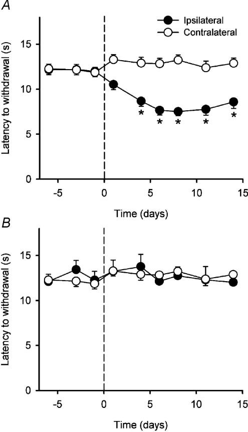
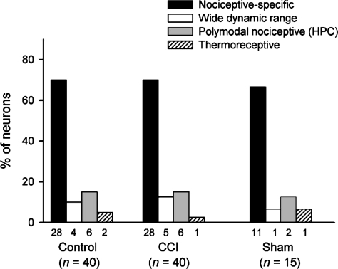
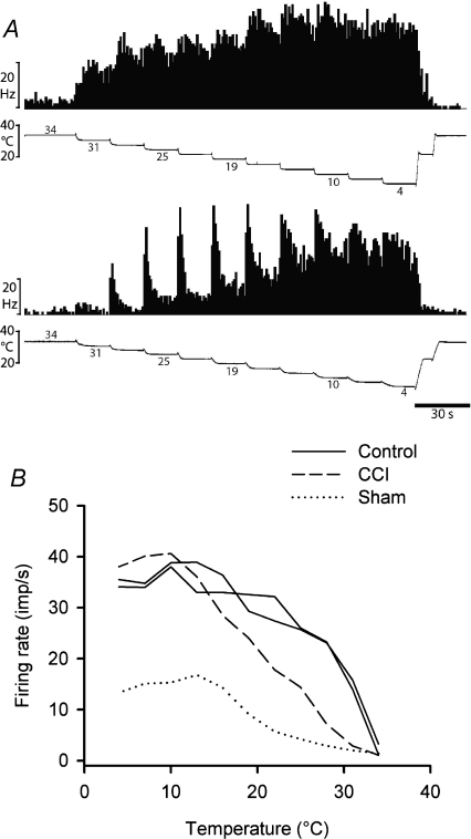
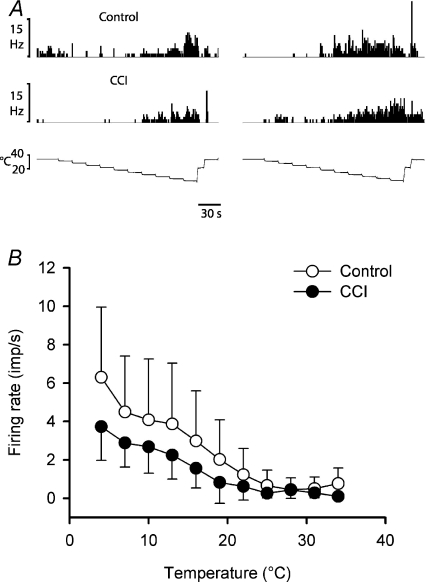
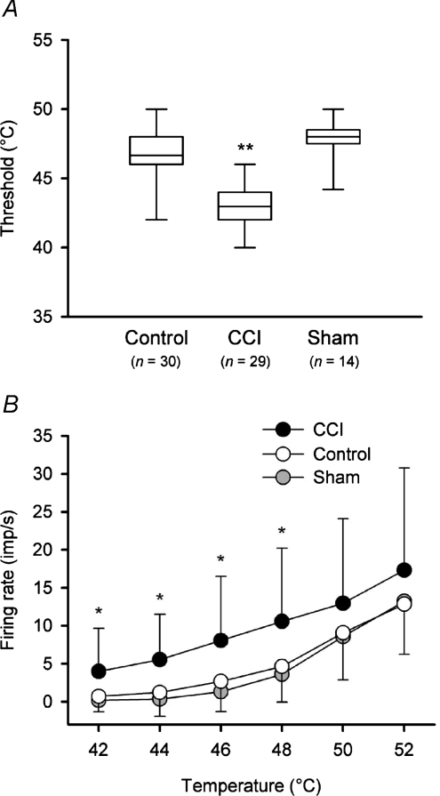
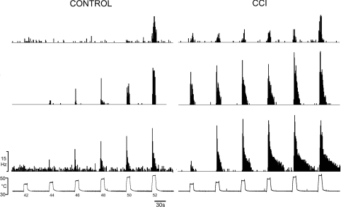
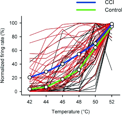
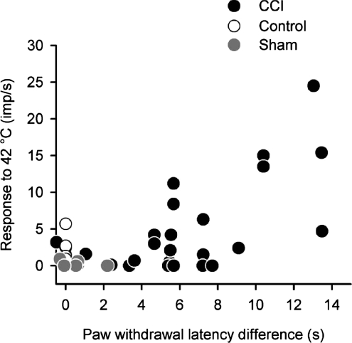
It has been proposed that spinal lamina I neurons with ascending axons that project to the midbrain play a crucial role in hyperalgesia. To test this hypothesis the quantitative properties of lamina I spinoparabrachial neurons in the chronic constriction injury (CCI) model of neuropathic pain were compared to those of unoperated and sham-operated controls. Behavioural testing showed that animals with a CCI exhibited heat hyperalgesia within 4 days of the injury, and this hyperalgesia persisted throughout the 14-day post-operative testing period. In the CCI, nociceptive lamina I spinoparabrachial neurons had heat thresholds that were significantly lower than controls (43.0 +/- 2.8 degrees C vs. 46.7 +/- 2.6 degrees C; P < 10(-4), ANOVA). Nociceptive lamina I spinoparabrachial neurons were also significantly more responsive to graded heat stimuli in the CCI, compared to controls (P < 0.02, 2-factor repeated-measures ANOVA), and increased after-discharges were also observed. Furthermore, the heat-evoked stimulus-response functions of lamina I spinoparabrachial neurons in CCI animals co-varied significantly (P < 0.03, ANCOVA) with the amplitude of heat hyperalgesia determined behaviourally. Taken together these results are consistent with the hypothesis that lamina I spinoparabrachial neurons have an important mechanistic role in the pathophysiology of neuropathic pain.


Figures

Figure 1
Figure 1. Identification of lamina I spinoparabrachial neurons in vivo
A, pair of traces showing 1-for-1 following of a train of 6 antidromic electrical stimuli (45 μA, 2 ms, 250 Hz; dots) delivered from the middle stimulating electrode in the contralateral parabrachial nucleus. B, collision of the first antidromic impulse in a train of 3 (150 Hz, upper trace) when an orthodromic impulse (asterisk, lower trace) occurred within the critical interval. The arrow indicates the point at which the first antidromic response should have occurred. C, photomicrograph of a frozen section stained with thionin showing the tracks of the stimulating electrodes (asterisks) in the contralateral parabrachial nucleus. Bar is 0.4 mm. bc, brachium conjunctivum; KF, Kölliker–Fuse nucleus; LC, locus coeruleus; PBel, external lateral subnucleus of the parabrachial area; PBil, internal lateral nucleus of the parabrachial area; PBm, medial subnucleus of the parabrachial area. D, photomicrograph of the contralateral spinal dorsal horn at the level of the 3rd lumbar segment. An arrowhead marks the position of an electrolytic microlesion that was made at the recording site of the cell shown in A and B. Dorsal is up, lateral is left. Bar is 0.2 mm.
Figure 2
Figure 2. Time course of the development of the behavioural effects of the CCI
A, mean (± 1 s.e.m.) paw withdrawal latencies to radiant heat stimulation of the ipsilateral and contralateral hindpaws in the CCI animals (n= 33). The CCI was performed on day 0 (indicated by the dashed line) Asterisks indicate significant differences (P < 0.02; 2-factor repeated-measures ANOVA followed by Tukey's post-hoc test) between the ipsilateral and contralateral paws. B, mean (± 1 s.e.m.) paw withdrawal latencies to radiant heat stimulation of the ipsilateral and contralateral hindpaws in the sham-operated animals (n= 13). The sham operation was performed on day 0 (indicated by the dashed line).
Figure 3
Figure 3. Frequency distribution of different functional classes of lamina I spinoparabrachial neurons in control, CCI and sham-operated animals
Each neuron isolated was classified using cutaneous stimuli of different modalities (see Methods). The bars show the frequency distribution of the 4 classes of lamina I spinoparabrachial neurons in the three groups of animals studied. There were no significant differences between groups (P > 0.3, χ2 test).
Figure 4
Figure 4. Stimulus encoding by lamina I spinoparabrachial COOL neurons
A, peristimulus time histograms from two cooling-specific neurons showing their responses to graded intensity cooling stimuli. The upper pair of records was from an unoperated control animal and the lower pair was from an animal that had received a CCI. B, stimulus–response curves of all 4 cooling-specific neurons isolated in the current study.
Figure 5
Figure 5. Encoding of cool and cold temperatures by HPC lamina I spinoparabrachial neurons
A, individual histogram responses from 4 HPC neurons (2 control, 2 CCI) to the standard cold stimulus sequence. B, mean (± 1 s.d.) stimulus–response curves to cold stimuli for HPC neurons in controls and CCI animals (n= 6 in each group). There was no significant difference between groups when the stimulus–response curves were compared (P > 0.2, 2-factor ANOVA).
Figure 7
Figure 7. Quantitative differences in neuronal heat encoding in animals with a CCI
A, box plots of heat thresholds of nociceptive neurons in each of the control, CCI and sham-operated groups. The horizontal line within the box is the median value, the box boundaries are the 25th and 75th percentiles and the bars indicate the data range. Thresholds were significantly lower in animals with a CCI (P < 10−4, ANOVA; asterisks) compared to both control and sham-operated rats. B, stimulus–response curves (mean ± 1 s.d.) of heat encoding by neurons in controls, CCI rats and sham-operated animals. Neurons in animals with a CCI were significantly more responsive to temperatures in the range 42–48°C (P < 0.003; 2-factor ANOVA followed by Tukey's post hoc test; asterisks).
Figure 6
Figure 6. Heat responsiveness of nociceptive lamina I spinoparabrachial neurons in control and neuropathic rats
Peristimulus time histograms of the discharge of 6 different lamina I spinoparabrachial neurons (3 control, 3 CCI) in response to graded heat stimulation. For each group of neurons (control and CCI), the middle histogram is from the cell whose maximum firing rate was the median of all of the neurons in that group; the top histogram is from the neuron whose maximum response was the 25th percentile of the population, and the bottom histogram is from the 75th percentile neuron. As can be seen, thresholds were lower in neurons recorded in CCI rats and suprathreshold responsiveness and after-discharge were also greater in those cells.
Figure 8
Figure 8. Normalized heat-evoked responses from nociceptive lamina I spinoparabrachial neurons
The individual stimulus–response functions of nociceptive lamina I spinoparabrachial neurons have been plotted, normalized to the maximal discharge of each cell. Red lines are from neurons in animals with a CCI and black lines are from neurons in unoperated controls. Population means are shown with thick blue (CCI) and green (control) lines. The discharge of neurons in animals with a CCI was significantly greater than that of controls at temperatures of 42–48°C (P < 0.002, general linear model).
Figure 9
Figure 9. Relationship between lamina I spinoparabrachial activity and behaviour
The discharge evoked by a 10 s duration 42°C heat stimulus has been plotted against the difference in heat-evoked withdrawal latency between the ipsilateral and contralateral hindpaws. Paw withdrawal latency difference was a significant co-variant in the heat-evoked stimulus–response curves of animals with a CCI when compared to sham-operated controls (P < 0.03, ANCOVA). As withdrawal latency differences for unoperated controls were not measured they have been assigned to zero for convenience.

References

    1. Ahlgren SC, White DM, Levine JD. Increased responsiveness of sensory neurons in the saphenous nerve of the streptozotocin-diabetic rat. J Neurophysiol. 1992;68:2077–2085. - PubMed
    1. Andrew D, Greenspan JD. Modality-specific hyper-responsivity of regenerated cat cutaneous nociceptors. J Physiol. 1999;516:897–906. - PMC - PubMed
    1. Andrew D, Craig AD. Quantitative responses of spinothalamic lamina I neurones to graded mechanical stimulation in the cat. J Physiol. 2002;545:913–931. - PMC - PubMed
    1. Attal N, Jazat F, Kayser V, Guilbaud G. Further evidence for ‘pain-related’ behaviours in a model of unilateral peripheral neuropathy. Pain. 1990;41:235–251. - PubMed
    1. Balasubramanyan S, Stemkowski PL, Stebbing MJ, Smith PA. Sciatic chronic constriction produces cell-type-specific changes in the electrophysiological properties of rat substantia gelatinosa neurons. J Neurophysiol. 2006;96:579–590. - PubMed

Publication types