Review
. 2009 May 1;587(Pt 9):1903-15.
doi: 10.1113/jphysiol.2008.167767. Epub 2009 Mar 16.

Dynamic integration of subplate neurons into the cortical barrel field circuitry during postnatal development in the Golli-tau-eGFP (GTE) mouse

Affiliations
Review

Dynamic integration of subplate neurons into the cortical barrel field circuitry during postnatal development in the Golli-tau-eGFP (GTE) mouse

Maria Carmen Piñon et al. J Physiol. .

Abstract

In the Golli-tau-eGFP (GTE) transgenic mouse the reporter gene expression is largely confined to the layer of subplate neurons (SPn), providing an opportunity to study their intracortical and extracortical projections. In this study, we examined the thalamic afferents and layer IV neuron patterning in relation to the SPn neurites in the developing barrel cortex in GTE mouse at ages embryonic day 17 (E17) to postnatal day 14 (P14). Serotonin transporter immunohistochemistry or cytochrome oxydase histochemistry was used to reveal thalamic afferent patterning. Bizbenzimide staining identified the developing cytoarchitecture in coronal and tangential sections of GTE brains. Enhanced green fluorescent protein (GFP)-labelled neurites and thalamic afferents were both initially diffusely present in layer IV but by P4-P6 both assumed the characteristic periphery-related pattern and became restricted to the barrel hollows. This pattern gradually changed and by P10 the GFP-labelled neurites largely accumulated at the layer IV-V boundary within the barrel septa whereas thalamic afferents remained in the hollows. To investigate whether this reorganisation is dependent on sensory input, the whiskers of row 'a' or 'c' were removed at P0 or P5 and the organisation of GFP-labelled neurites in the barrel cortex was examined at P6 or P10. In the contralateral region corresponding to row 'a' or 'c' the lack of hollow to septa rearrangement of the GFP-labelled neurites was observed after P0 row removal at P10 but not at P6. Our findings suggest a dynamic, sensory periphery-dependent integration of SPn neurites into the primary somatosensory cortex during the period of barrel formation.


Figures
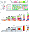
Figure 1
Figure 1. The Golli-tau-eGFP mouse as a model to study subplate neurite integration into the barrel cortex during development
A, schematic of the GTE construct (adopted from Jacobs et al. 2007). B, schematic diagram of the sensory pathway from the whiskers to the contralateral barrel cortex. C, schematic diagram of a small sector from a tangential section through a P0 barrel cortex with homogeneous distribution of layer IV neurons (blue), thalamic afferents (red) and GFP neurites (green). At P6, the thalamic projections assume the characteristic periphery-related pattern and target the barrel hallows and the layer IV neurons cluster at the septa (diagram based on Molnár & Molnár, 2006). There are three possible scenarios of the GFP neurite arrangement: (i) non-specific distribution; (ii) intra-barrel patterning; and (iii) inter-barrel patterning. D, schematic representation of the thalamic fibre ingrowth in the mouse primary somatosensory cortex. At embryonic day 11 (E11), the first postmitotic cells migrate to the outer edge of the cerebral wall to form the preplate (PP), which is subsequently split into the marginal zone (MZ, the future layer I) and the subplate (SP) by the arrival of neurons in the true cortical plate (CP). When thalamic axons (red lines) arrive to the cortex at E15, only a densely packed cortical plate is present. The axons start to accumulate in SP, although some axons and side branches penetrate the deep part of the cortical plate (DCP). During the early postnatal period (P0), most thalamic fibres invade the CP and layers V and VI. Thalamic axons assume their characteristic periphery-related pattern and impose a barrel arrangement on cortical layer IV neurons. E, optical recording with voltage-sensitive dyes revealed that thalamocortical projections elicit sustained depolarization in the SP at E18–19. This depolarization spreads to almost the entire thickness of the cortex shortly after birth and concentrate by the end of the first postnatal week (P8) at the periphery-related clusters in layer IV. IZ, intermediate zone; SVZ, subventricular zone; VZ, ventricular zone; WM, white matter. Adapted and modified from Molnár & Hannan (2000) and López-Bendito & Molnár (2003).
Figure 3
Figure 3. Fluorescence micrographs of coronal sections through the developing barrel cortex of the GTE mouse at ages E17–P14 (AH) and thalamic axon segregation pattern in relation to the GFP-labelled neurites in the GTE mouse from P6 to P14 (IQ)
GFP neurites are shown in green, thalamic afferents (labelled with 5HTT immunohistochemistry) in red and the bisbenzimide nuclear counterstain shown in blue. A–C, GFP-labelled neurites extend radially towards the pial surface from cell bodies situated in SP and to lesser extent in cortical plate (layer VI). By P4 and P6 GFP-positive fibres cluster in layer IV, but subsequently this pattern is changed by the retraction of the GFP-labelled neurites from the hollows of layer IV. Filled arrowheads in D and E indicate intra-barrel GFP densities; arrows in F indicate barrel septa; open arrowheads in G and H indicate inter-barrel GFP concentration. I, L and O, thalamic axons were visualized by serotonin immunohistochemistry for serotonin transporter (5HTT) which show a clear segregation in a periphery-related pattern at P6, P10 and P14. To relate the subplate neurites in relation to the thalamic afferents, coronal sections through the barrel cortex of a GTE mouse were immunostained for 5HTT. At P6, the GFP neurite innervation from the subplate is confined to the barrels (filled arrowheads in J) similarly to the serotonin transporter immunoreactivity pattern (I) but by P10 and P14 the GFP neurite innervation and the serotonin immunohistochemistry pattern do not coincide with each other (L, M, O and P). GFP-labelled fibres are confined to the septa at P10 and P14 (open arrowheads in M and P). Abbreviations: mz marginal zone; cp, cortical plate; sp, subplate; wm, white matter; IV, layer IV; hp, hippocampus. Scale bar, 100 μm (A–C, I–Q) and 200 μm (D–H).
Figure 2
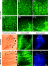
Figure 2. Age specific rearrangement of the GFP positive neurite and the effect of whisker removal at birth
AF, fluorescence micrographs of the barrel cortex in the tangential plane of the Golli-tau-eGFP (GTE) mouse revealed a changing pattern of GFP-labelled neurite distribution in layer IV between P2–14. The initially homogeneous distribution (A–C) changes to hollow (P6, C), then to septa (P10–14, D–F). GL, the effect of neonatal whisker removal on the GFP neurite patterning in the P10 barrel cortex. Bright field (G and J) and fluorescence micrographs (H, I, K and L) comparing the two barrel cortices in the tangential sections of a Golli-tau-eGFP mouse at P10. G, H and I are from the control side (ipsilateral hemisphere to the whisker removal) and J, K and L are from the same mouse contralateral to the whisker ablation from row ‘a’ at P0. Dashed rings encircle the row ‘a’ region of the barrel cortex. Scale bar for AF, 200 μm; for GL, 300 μm.
Figure 4
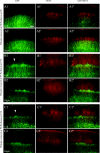
Figure 4. The effect of the timing of row ’c’ whisker removal on the GFP neurite patterning in the somatosensory cortex of the Golli-tau-eGFP mouse
Three different protocols were investigated: row ‘c’ was removed at P0 and studied at P6 (A), row ‘c’ was removed at P5 and studied at P10 (B) and row ‘c’ was removed at P0 and studied at P10 (C). Fluorescence micrographs illustrate the GFP (left), 5HTT (middle) and GFP and 5HTT merged patterning (right column) on coronal sections at P6 (A) and P10 (B and C) from the contralateral (labelled with 1) and ipsilateral (labelled with 2) side to the whisker removal. Arrowheads in B1 and C1 point to the area with remaining GFP neurite innervation towards the barrel hollow, corresponding to the region innervated by the removed whiskers row ‘c’. There is no clear change in the GFP neurite patterning at P6 after P0 whisker ablation. This further supports the observations (illustrated in Figs 1 and 2), that the GFP innervation patterning on the barrel cortex changes from an intra-barrel to an inter-barrel pattern after P6. Scale bar, 200 μm.

References

    1. Agmon A, Yang LT, Jones EG, O’Dowd DK. Topological precision in the thalamic projection to neonatal mouse barrel cortex. J Neurosci. 1995;15:549–561. - PMC - PubMed
    1. Allendoerfer KL, Shatz CJ. The subplate, a transient neocortical structure: its role in the development of connections between the thalamus and cortex. Annu Rev Neurosci. 1994;17:185–218. - PubMed
    1. Antonini A, Shatz CJ. Relation between putative transmitter phenotypes and connectivity of subplate neurons during cerebral cortical development. Eur J Neurosci. 1990;2:744–761. - PubMed
    1. Arber S. Subplate neurons: bridging the gap to function in the cortex. Trends Neurosci. 2004;27:111–113. - PubMed
    1. Aye L, Piñon MC, Hoerder A, Jacobs E, Campagnoni A, Molnár Z. Properties of layer 6a and subplate neurons in the Golli Tau eGFP (GTE) mouse. FENS Abstr. 2006;3:A196.

Publication types