. 2009 Mar 23;184(6):777-84.
doi: 10.1083/jcb.200811102. Epub 2009 Mar 16.

Wac: a new Augmin subunit required for chromosome alignment but not for acentrosomal microtubule assembly in female meiosis

Affiliations

Wac: a new Augmin subunit required for chromosome alignment but not for acentrosomal microtubule assembly in female meiosis

Ana M Meireles et al. J Cell Biol. .

Abstract

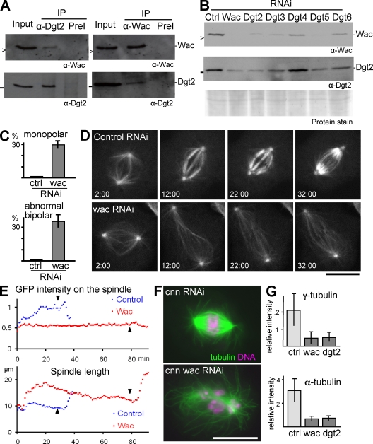
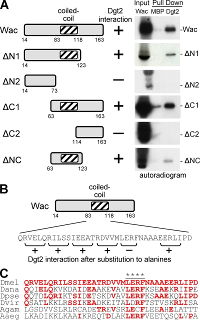
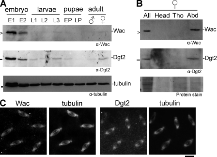
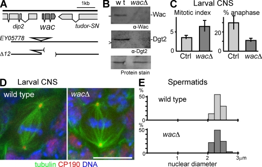
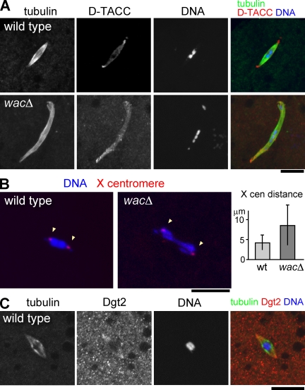
The bipolar spindle forms without centrosomes naturally in female meiosis and by experimental manipulation in mitosis. Augmin is a recently discovered protein complex required for centrosome-independent microtubule generation within the spindle in Drosophila melanogaster cultured cells. Five subunits of Augmin have been identified so far, but neither their organization within the complex nor their role in developing organisms is known. In this study, we report a new Augmin subunit, wee Augmin component (Wac). Wac directly interacts with another Augmin subunit, Dgt2, via its coiled-coil domain. Wac depletion in cultured cells, especially without functional centrosomes, causes severe defects in spindle assembly. We found that a wac deletion mutant is viable but female sterile and shows only a mild impact on somatic mitosis. Unexpectedly, mutant female meiosis showed robust microtubule assembly of the acentrosomal spindle but frequent chromosome misalignment. For the first time, this study establishes the role of an Augmin subunit in developing organisms and provides an insight into the architecture of the complex.


Figures

Figure 1.
Figure 1.
Wac is an integral component of Augmin. (A) Wac and Dgt2 reciprocally coimmunoprecipitated. Wac or Dgt2 was immunoprecipitated from the soluble fraction (input) of S2 cell extract using preimmune sera (PreI) as a control (Ctrl) and immunoblotted for these two proteins. > and − indicate 16.5 kD and 25 kD, respectively. (B) Decreased amounts of Wac and Dgt2 after depletion of Augmin subunits (Dgt2–6). (C) The frequencies of monopolar spindles and abnormal bipolar spindles, typically with reduced microtubule density, after 5 d of dsRNA treatment. (D) Time sequences of mitotic progression in S2 cells expressing GFP–α-tubulin after 3 d of dsRNA incubation. Time (minutes:seconds) after the nuclear envelope breakdown is shown. (E) Change in the GFP–α-tubulin signal intensity on the spindles shown in B and change in the spindle length. The intensity was normalized against signals around the poles at prophase. The arrowheads indicate anaphase onset. (F) Typical spindle morphologies of single centrosomin (Cnn) depletion and of codepletion with Wac. (G) The γ- and α-tubulin signal intensity on the spindle relative to the poles after RNAi. IP, immunoprecipitation. Error bars indicate SD. Bars, 10 µm.
Figure 2.
Figure 2.
Wac directly interacts with Dgt2 via its coiled-coil region. (A) A series of Wac truncations were translated in vitro in reticulocyte lysate in the presence of [35S]-methionine (input), and interaction with bacterially produced MBP-Dgt2 (Dgt2) or MBP were tested by pull-down. (B) Consecutive residues within the coiled-coil region of Wac were systematically replaced with alanines, and the interaction with Dgt2 was tested. (C) A sequence comparison of Wac homologues among insects. Red letters indicate residues identical to those in D. melanogaster, and the asterisks indicate residues essential for the interaction. Dmel, D. melanogaster; Dana, Drosophila ananassae; Dpse, Drosophila pseudoobscura; Dvir, Drosophila virilis; Agam, Anopheles gambiae; Aaeg, Aedes aegypti.
Figure 3.
Figure 3.
Expression and localization of Wac and Dgt2 in developing flies. (A) Expression of Wac and Dgt2 proteins during development. E1 and E2 indicate 0–4-h-old and 4–20-h-old embryos, respectively; L1, L2, and L3 indicate first, second, and third instar larvae, respectively; EP and LP indicate early and late darkened pupae, respectively; >, −, and • indicate 16.5 kD, 25 kD, and 55 kD, respectively. (B) Expression of Wac and Dgt2 proteins in adult females. All, whole body of adult females; Tho, thorax; Abd, abdoman. (C) Immunolocalization of Wac and Dgt2 to spindle microtubules in syncytial embryos. Bar, 10 µm.
Figure 4.
Figure 4.
Wac is dispensable for somatic mitosis and male meiosis. (A) The genomic organization of the wacΔ mutant. Parentheses represent the deleted region. (B) Immunoblots showing the absence of Wac and Dgt2 proteins in wacΔ adult females. wt, wild type. (C) Increased preanaphase stages in wacΔ. The mitotic index and the frequency of anaphase among mitotic cells in orcein-stained brain squashes with the SD (n = 4). The differences are significant between the homozygous mutant and the control (Ctrl) sibling heterozygotes (P < 0.01). (D) Immunostaining of whole-mount wild-type and wacΔ larval neuroblasts. CP190 is a centrosomal protein. (E) The diameter of spermatid nuclei at the onion stage from wild type and the wacΔ mutant. The distribution is not significantly different between the wild type and mutant (by χ2 test; n > 100). Error bars indicate SD. Bar, 10 µm.
Figure 5.
Figure 5.
Wac is not required for spindle microtubule assembly but for chromosome alignment in female meiosis. (A) Metaphase I spindle in nonactivated matured oocytes. Chromosomes misalign asymmetrically within a robust bipolar spindle in wacΔ. (B) Location of an X chromosome centromeric satellite at metaphase I determined by FISH. The mean distance between homologous centromeres significantly increased in wacΔ (P < 0.01). cen, centromere; wt, wild type. (C) Dgt2 accumulates at the polar regions of the acentrosomal metaphase I spindle in wild type. Error bars indicate SD. Bars, 10 µm.

References

    1. Ashburner M., Golic K.G., Hawley R.S. 2005. Drosophila: A Laboratory Handbook. Cold Spring Harbor Laboratory Press, Cold Spring Harbor, NY: 1409
    1. Basto R., Lau J., Vinogradova T., Gardiol A., Woods C.G., Khodjakov A., Raff J.W. 2006. Flies without centrioles.Cell. 125:1375–1386 - PubMed
    1. Cullen C.F., Ohkura H. 2001. Msps protein is localized to acentrosomal poles to ensure bipolarity of Drosophila meiotic spindles.Nat. Cell Biol. 3:637–642 - PubMed
    1. Cullen C.F., Deák P., Glover D.M., Ohkura H. 1999. mini spindles: a gene encoding a conserved microtubule-associated protein required for the integrity of the mitotic spindle in Drosophila.J. Cell Biol. 146:1005–1018 - PMC - PubMed
    1. Cullen C.F., Brittle A.L., Ito T., Ohkura H. 2005. The conserved kinase NHK-1 is essential for mitotic progression and unifying acentrosomal meiotic spindles in Drosophila melanogaster.J. Cell Biol. 171:593–602 - PMC - PubMed

Publication types

MeSH terms