. 2009 Mar 31;106(13):5318-23.
doi: 10.1073/pnas.0811713106. Epub 2009 Mar 16.

Resistance to CCR5 inhibitors caused by sequence changes in the fusion peptide of HIV-1 gp41

Affiliations

Resistance to CCR5 inhibitors caused by sequence changes in the fusion peptide of HIV-1 gp41

Cleo G Anastassopoulou et al. Proc Natl Acad Sci U S A. .

Abstract

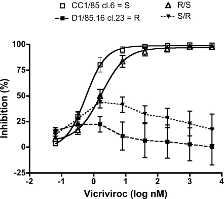
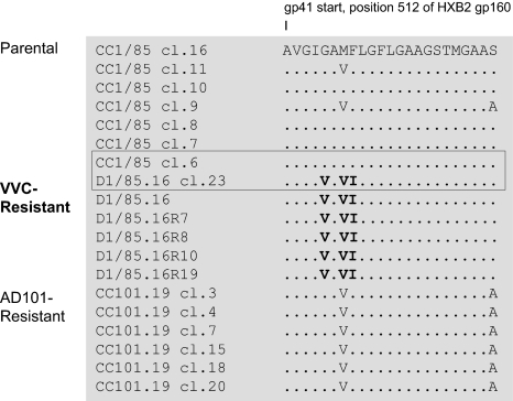
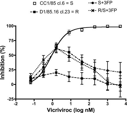
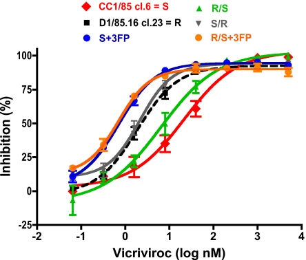
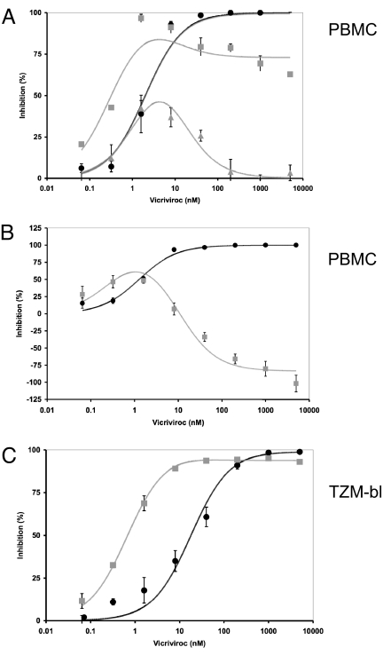
We have investigated the mechanism of resistance of a HIV type 1 (HIV-1) R5 primary isolate, D1/85.16, to the small molecule CCR5 inhibitor, vicriviroc (VVC). Unlike other viruses resistant to this class of compound, D1/85.16 lacks sequence changes in the V3 region of the gp120 surface glycoprotein. Inspection of env sequences from D1/85.16 compared with those derived from the parental, inhibitor-sensitive virus, CC1/85, revealed a cluster of 3 conservative changes in the fusion peptide (FP) of the gp41 transmembrane glycoprotein that tracked with the resistance phenotype. Studies with engineered Env-chimeric and point-substituted viruses confirmed that these 3 FP residues were substantially responsible for VVC resistance without altering coreceptor usage, as assessed in both peripheral blood mononuclear cells and the TZM-bl cell line. VVC resistance is manifested differently in the 2 cell types, and there are assay-dependent complexities to the dose-response curves for the engineered resistant viruses. To explain them, we created a model for resistance and generated theoretical VVC inhibition curves that closely mimic the experimental data for the resistant viruses. The basis for the model is the existence of distinct forms of CCR5, with varying affinities for small molecule CCR5 inhibitors that are presumed to be present in different proportions on different cell types, and are used selectively by resistant HIV-1 variants when ligated with an inhibitor. Together, the experimental results and theoretical model may help understand how HIV-1 uses CCR5 to enter target cells under various conditions.


Conflict of interest statement

The authors declare no conflict of interest.

Figures

Fig. 1.
Fig. 1.
VVC resistance maps to gp41. Clonal chimeric viruses R/S and S/R (Table S1) bearing gp120 and gp41 subunits derived either from the parental, VVC-sensitive clone S, or the VVC-resistant clone R were tested for VVC sensitivity in a multicycle PBMC-based replication assay measuring p24 production 7 days postinfection. The data shown are mean values derived from 7 to 15 independent experiments ± SEM.
Fig. 2.
Fig. 2.
Alignment of N-terminal gp41 sequences from VVC-sensitive and VVC-resistant viruses. The first 23 amino acid residues from the gp41 N terminus are shown for 7 clones from the CC1/85 parental virus, 6 VVC-resistant isolates or clones based on D1/85.16, and 6 clones from the AD101-resistant CC101.19 isolate. The sequences are recorded relative to that of CC1/85 cl.16 (top line), with dashes indicating amino acid sequence identity. Amino acid numbering is based on HXB2 Env, with the first residue of gp41 at position 512. The 3 conservative substitutions of hydrophobic residues, G516V, M518V, and F519I, in the FP region of resistant viruses are highlighted in bold. Among the 7 parental clones, S (GenBank accession no. AY357338) is the most closely related to R; these 2 clones, which were used for subsequent genetic studies, are boxed. The env sequences of the D1/85.16-derived viruses have been deposited in GenBank (accession nos. FJ713453–FJ713458).
Fig. 3.
Fig. 3.
The 3 gp41 FP amino acid substitutions confer resistance to VVC. Site-directed mutagenesis was performed to introduce the 3 changes (G516V, M518V, and F519I) into the VVC-sensitive parental clone S and the R/S chimera (Table S1). The engineered mutant viruses, S + 3FP and R/S + 3FP, were then tested for VVC sensitivity in a multicycle PBMC-based replication assay measuring p24 production 7 days postinfection. For comparison, S and the VVC-resistant clone R were also tested. In the same experiments, the R/S and S/R chimeras behaved comparably with S and R, respectively, but the curves are not shown, for clarity. The depicted results are the average of 6–10 independent experiments, with the error bars indicating the SEM.
Fig. 4.
Fig. 4.
VVC resistance is manifested differently in a cell line-based assay. The same infectious, clonal viruses studied in PBMC were tested in TZM-bl cells with a luciferase reporter gene endpoint. The VVC inhibition curves (dashed black line for the resistant virus R) asymptote to a plateau that is high, but consistently lower (upper limit of 95% C.I. <100%) for the resistant than the sensitive viruses. The data shown are the average of 3 independent experiments. Resistance-related parameters derived from these experiments are summarized in Table 1.
Fig. 5.
Fig. 5.
Fitting of model function to empirical data from PBMC and TZM-bl cell assays. The relative inhibition of virus infectivity is expressed in percentage on the y axis as a function of the VVC concentration in nM on the x axis. The black circles represent data for wild-type virus (S); data for the VVC-resistant S + 3FP mutant are shown as gray squares and triangles (error bars, SEM in each case). The theoretical curves derived from the model are shown as continuous lines in the shades of the dataset each is fitted to. (A) Inhibition of the S + 3FP (gray) and S (black) viruses in PBMC from 2 different donor pools. The resistant virus is partially or negligibly inhibited at the highest VVC concentrations. Datasets from other donor pools fall between these profiles when plotted similarly. (B) An example of enhancement of S + 3FP virus infection (gray) at the highest VVC concentration is contrasted with inhibition of S virus (black) on PBMC from different donor pools. Different relative amounts of the CCR5-A and CCR5-B modulate the different levels of inhibition seen at the highest VVC concentrations in the different experiments. (C) Inhibition of TZM-bl cell infection by S + 3FP (gray) and S (black). The model fits the data well when the VVC affinities of CCR5-A and CCR5-B are assumed to differ by >37-fold, a value that was predetermined by measuring the IC50 values for the respective viruses on TZM-bl cells. The small residual infectivity of S + 3FP at the highest VVC concentrations results from an excess of CCR5-B (high-affinity for VVC) over CCR5-A (lower affinity for VVC). This excess would be ≈20-fold if the efficiency of S + 3FP viral entry were the same for free CCR5-B as for VVC-CCR5-A complexes.

References

    1. Kuhmann SE, Hartley O. Targeting chemokine receptors in HIV: A status report. Annu Rev Pharmacol Toxicol. 2008;48:425–461. - PubMed
    1. Moore JP, Kitchen SG, Pugach P, Zack JA. The CCR5 and CXCR4 coreceptors-central to understanding the transmission and pathogenesis of human immunodeficiency virus type 1 infection. AIDS Res Hum Retroviruses. 2004;20:111–126. - PubMed
    1. Westby M, et al. Emergence of CXCR4-using human immunodeficiency virus type 1 (HIV-1) variants in a minority of HIV-1-infected patients following treatment with the CCR5 antagonist maraviroc is from a pretreatment CXCR4-using virus reservoir. J Virol. 2006;80:4909–4920. - PMC - PubMed
    1. Westby M, et al. Reduced maximal inhibition in phenotypic susceptibility assays indicates that viral strains resistant to the CCR5 antagonist maraviroc utilize inhibitor-bound receptor for entry. J Virol. 2007;81:2359–2371. - PMC - PubMed
    1. Pugach P, et al. HIV-1 clones resistant to a small molecule CCR5 inhibitor use the inhibitor-bound form of CCR5 for entry. Virology. 2007;361:212–228. - PMC - PubMed

Publication types