. 2009 Mar 31;106(13):5105-10.
doi: 10.1073/pnas.0900026106. Epub 2009 Mar 16.

Designed amphiphilic peptide forms stable nanoweb, slowly releases encapsulated hydrophobic drug, and accelerates animal hemostasis

Affiliations

Designed amphiphilic peptide forms stable nanoweb, slowly releases encapsulated hydrophobic drug, and accelerates animal hemostasis

Liping Ruan et al. Proc Natl Acad Sci U S A. .

Abstract

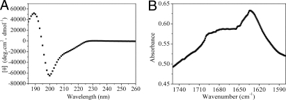
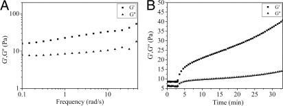
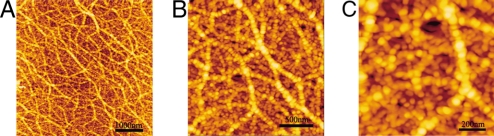
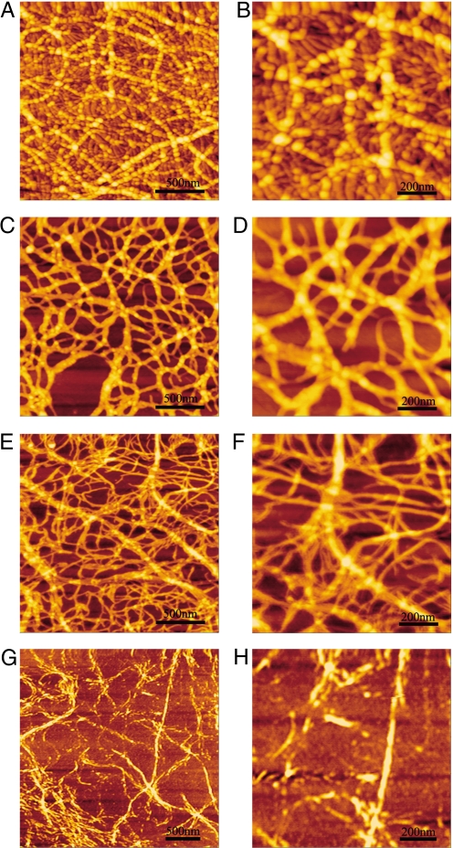
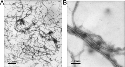
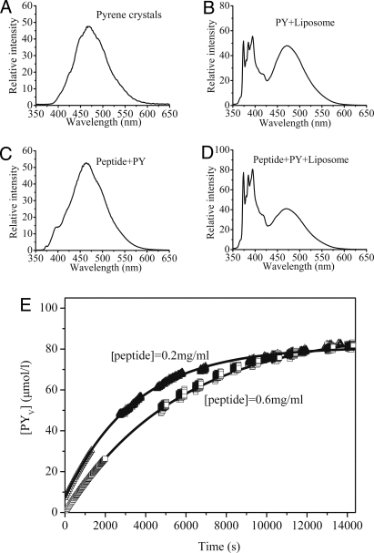
How do you design a peptide building block to make 2-dimentional nanowebs and 3-dimensional fibrous mats? This question has not been addressed with peptide self-assembling nanomaterials. This article describes a designed 9-residue peptide, N-Pro-Ser-Phe-Cys-Phe-Lys-Phe-Glu-Pro-C, which creates a strong fishnet-like nanostructure depending on the peptide concentrations and mechanical disruptions. This peptide is intramolecularly amphiphilic because of a single pair of ionic residues, Lys and Glu, at one end and nonionic residues, Phe, Cys, and Phe, at the other end. Circular dichroism and Fourier transform infrared spectroscopy analysis demonstrated that this peptide adopts stable beta-turn and beta-sheet structures and self-assembles into hierarchically arranged supramolecular aggregates in a concentration-dependent fashion, demonstrated by atomic force microscopy and electron microscopy. At high concentrations, the peptide dominantly self-assembled into globular aggregates that were extensively connected with each other to form "beads-on-a-thread" type nanofibers. These long nanofibers were extensively branched and overlapped to form a self-healing peptide hydrogel consisting of >99% water. This peptide can encapsulate the hydrophobic model drug pyrene and slowly release pyrene from coated microcrystals to liposomes. It can effectively stop animal bleeding within 30 s. We proposed a plausible model to interpret the intramolecular amphiphilic self-assembly process and suggest its importance for the future development of new biomaterials for drug delivery and regenerative medicine.


Conflict of interest statement

The authors declare no conflict of interest.

Figures

Fig. 1.
Fig. 1.
Secondary structures of the peptide. (A) CD spectrum of the peptide (200 μM) at 25 °C in the Milli-Q water. The spectrum shows a negative maximum ≈200 nm ([θ]200 = −65,000° per cm2·dmol−1) and a positive maximum near 189 nm ([θ]189 = 52,000° per cm2·dmol−1), the characteristics of a β-turn structure. (B) FTIR spectrum for the peptide at 10 mg/mL. The peptide has a peak centered at ≈1,633 cm−1, and this peak is attributed to the formation of sheet structure. The other two broad peaks are located at ≈1,675 and 1,696 cm−1, characteristics of the β-turn structure.
Fig. 2.
Fig. 2.
Rheology of the peptide solution at 10 mg/mL. (A) Frequency sweep (1-Pa shear stress) of 1% peptide at 25 °C. ■, storage moduli; ▴, loss moduli. The frequency sweep results measured at 25 °C revealed that storage modulus of the peptide (G′, a measure of the elastic response of the material) was larger than loss modulus (G″, a measure of the viscous response) over all measured frequencies. (B) Restoration of gel as a function of time followed by the cessation of treatment for gel network destruction (1,000% strain at 6 Hz for 180 s). ■, storage moduli; ▴, loss moduli. The peptide showed its strong self-assembling ability to reform the gel quickly.
Fig. 3.
Fig. 3.
Typical AFM morphological images of the peptide deposited on mica. (A) Peptide dissolved in water at 1 mg/mL. The image shows beads-on-a-thread fibers in a branched network. These fibers are made of many globular aggregates lining up and stacking together. (B) Higher magnification of A; a staggered arrangement of globular aggregates is observed. (C) Higher magnification of B. The scales are marked in each panel.
Fig. 4.
Fig. 4.
AFM images at different peptide concentrations. (A) At 1 mg/mL, showing globular structure fibers and many small fibers in the background. (B) Higher magnification of A, showing details of the background with small fibers and small globular structures. (C and D) At 0.5 mg/mL, showing little globular structures along the fibers. (E and F) At 0.3 mg/mL, showing many small fibers and some wider fibers twisted together with the smaller ones. (G and H) At 0.1 mg/mL, showing many small fiber and narrow fibers. The scales are marked in each panel.
Fig. 5.
Fig. 5.
TEM images of the peptide at 0.2 mg/mL. (A) Low magnification (25,000-fold), showing high-density fiber networks. (B) Higher magnification (60,000-fold) for a single fiber, and the fibrils were twisted together to form fibers. The scales are marked in each panel.
Fig. 6.
Fig. 6.
Steady-state fluorescence emission spectra. (A–D) Solid pyrene crystals (A) and pyrene in soybean lecithin liposome vesicles (B) ([PY] = 9.6 × 10⁻⁵ M; [liposome] = 1.59 × 10⁻³ M). (C) Peptide–PY solution ([PY] = 1.98 × 10⁻³ M; [peptide] = 1.75 × 10⁻⁴ M), showing a spectrum similar to A. (D) Peptide–PY solution mixed with liposome vesicles ([PY] = 6.6 × 10⁻⁵M ; [peptide] = 5.83 × 10⁻⁶ M; [liposome] = 1.59 × 10− 3M), λex = 336 nm, showing spectrum similar to B. (E) Fluorescence curves for the release of molecular pyrene from pyrene microcrystals encapsulated in the peptide coating into bilayers of soybean lecithin liposome ([liposome] = 1.59 × 10⁻³ M). □ and ▵, pyrene transfer experiments carried out with peptide 06–PY and peptide 02–PY, respectively.

References

    1. Zhang S, Holmes T, Lockshin C, Rich A. Spontaneous assembly of a self-complementary oligopeptide to form a stable macroscopic membrane. Proc Natl Acad Sci USA. 1993;90:3334–3338. - PMC - PubMed
    1. Zhang S, Gelain F, Zhao X. Designer self-assembling peptide nanofiber scaffolds for 3D tissue cell cultures. Semin Cancer Biol. 2005;15:413–420. - PubMed
    1. Zhao X, Zhang S. Self-assembling nanopeptides become a type of biomaterial. Adv Polym Sci. 2006;203:145–170.
    1. Zhang S. Building from the bottom up. Materials Today. 2003;6:20–27.
    1. Yokoi H, Kinoshita T, Zhang S. Dynamic reassembly of peptide RADA16-I nanofiber scaffold. Proc Natl Acad Sci USA. 2005;102:8414–8419. - PMC - PubMed

Publication types

LinkOut - more resources