

Epitope characterization and variable region sequence of f1-40, a high-affinity monoclonal antibody to botulinum neurotoxin type a (Hall strain)
- PMID: 19290051
- PMCID: PMC2654115
- DOI: 10.1371/journal.pone.0004924
Epitope characterization and variable region sequence of f1-40, a high-affinity monoclonal antibody to botulinum neurotoxin type a (Hall strain)
Erratum in
- PLoS One. 2009;4(10). doi: 10.1371/annotation/47f9bc3f-7c79-453c-85e3-588bc3056951 doi: 10.1371/annotation/47f9bc3f-7c79-453c-85e3-588bc3056951
- PLoS One. 2009;4(10). doi: 10.1371/annotation/6359a6c2-90f8-4f7e-ad86-5bac5d62a7fe doi: 10.1371/annotation/6359a6c2-90f8-4f7e-ad86-5bac5d62a7fe
- PLoS One. 2009;4(10). doi: 10.1371/annotation/8d83a0a4-4dd0-4c57-ad94-304130f565e6 doi: 10.1371/annotation/8d83a0a4-4dd0-4c57-ad94-304130f565e6
- PLoS One. 2009;4(11). doi: 10.1371/annotation/6359a6c2-90f8-4f7e-ad86-5bac5d62a7fe doi: 10.1371/annotation/6359a6c2-90f8-4f7e-ad86-5bac5d62a7fe
Abstract
Background: Botulism, an often fatal neuroparalytic disease, is caused by botulinum neurotoxins (BoNT) which consist of a family of seven serotypes (A-H) produced by the anaerobic bacterium Clostridium botulinum. BoNT, considered the most potent biological toxin known, is a 150 kDa protein consisting of a 100 kDa heavy-chain (Hc) and a 50 kDa light-chain (Lc). F1-40 is a mouse-derived, IgG1 monoclonal antibody that binds the light chain of BoNT serotype A (BoNT/A) and is used in a sensitive immunoassay for toxin detection. We report the fine epitope mapping of F1-40 and the deduced amino acid sequence of the variable regions of the heavy and light chains of the antibody.
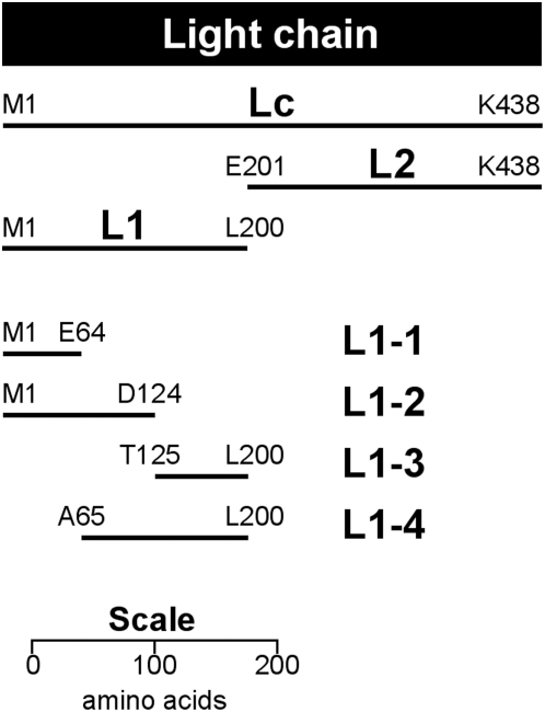
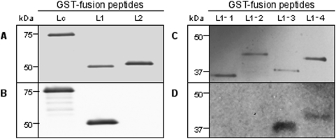
Methods and findings: To characterize the binding epitope of F1-40, three complementary experimental approaches were selected. Firstly, recombinant peptide fragments of BoNT/A light-chain were used in Western blots to identify the epitope domains. Secondly, a peptide phage-display library was used to identify the specific amino acid sequences. Thirdly, the three-dimensional structure of BoNT/A was examined in silico, and the amino acid sequences determined from the phage-display studies were mapped onto the three-dimensional structure in order to visualize the epitope. F1-40 was found to bind a peptide fragment of BoNT/A, designated L1-3, which spans from T125 to L200. The motif QPDRS was identified by phage-display, and was mapped to a region within L1-3. When the three amino acids Q138, P139 and D140 were all mutated to glycine, binding of F1-40 to the recombinant BoNT/A light chain peptide was abolished. Q-138, P-139 and D-140 form a loop on the external surface of BoNT/A, exposed to solvent and accessible to F1-40 binding.
Conclusions: The epitope of F1-40 was localized to a single exposed loop (ss4, ss5) on the Lc of BoNT. Furthermore amino acids Q138, P139 and D140 forming the tip of the loop appear critical for binding.
Conflict of interest statement
Figures
References
-
- Lamanna C. The most poisonous poison. Science. 1959;130:763–772. - PubMed
-
- Niwa K, Koyama K, Inoue S, Suzuki T, Hasegawa K, et al. Role of nontoxic components of serotype D botulinum toxin complex in permeation through a Caco-2 cell monolayer, a model for intestinal epithelium. FEMS Immunol Med Microbiol. 2007;49(3):346–352. - PubMed
-
- Montecucco C, Schiavo G. Tetanus and botulism neurotoxins: a new group of zinc proteases. Trends Biochem Sci. 1993;18(9):324–327. - PubMed
Publication types
MeSH terms
Substances
LinkOut - more resources
Full Text Sources
Other Literature Sources
Molecular Biology Databases
Research Materials
