. 2009;4(3):e4914.
doi: 10.1371/journal.pone.0004914. Epub 2009 Mar 17.

A conserved mechanism for control of human and mouse embryonic stem cell pluripotency and differentiation by shp2 tyrosine phosphatase

Affiliations

A conserved mechanism for control of human and mouse embryonic stem cell pluripotency and differentiation by shp2 tyrosine phosphatase

Dongmei Wu et al. PLoS One. 2009.

Abstract

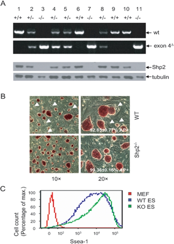
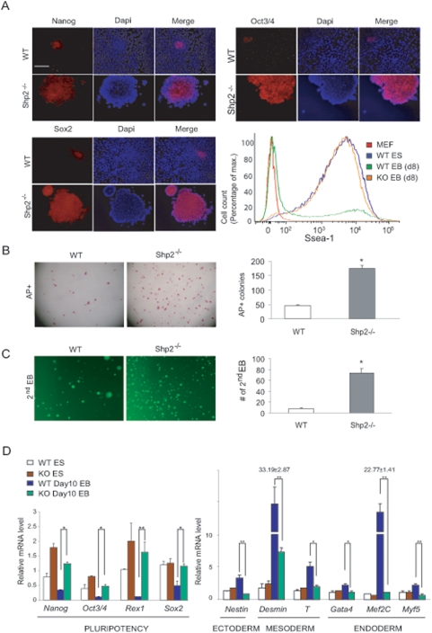
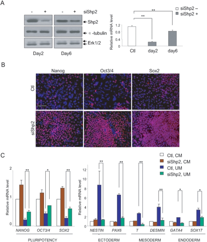
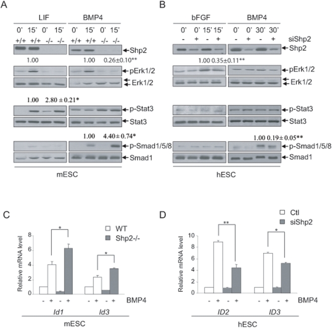
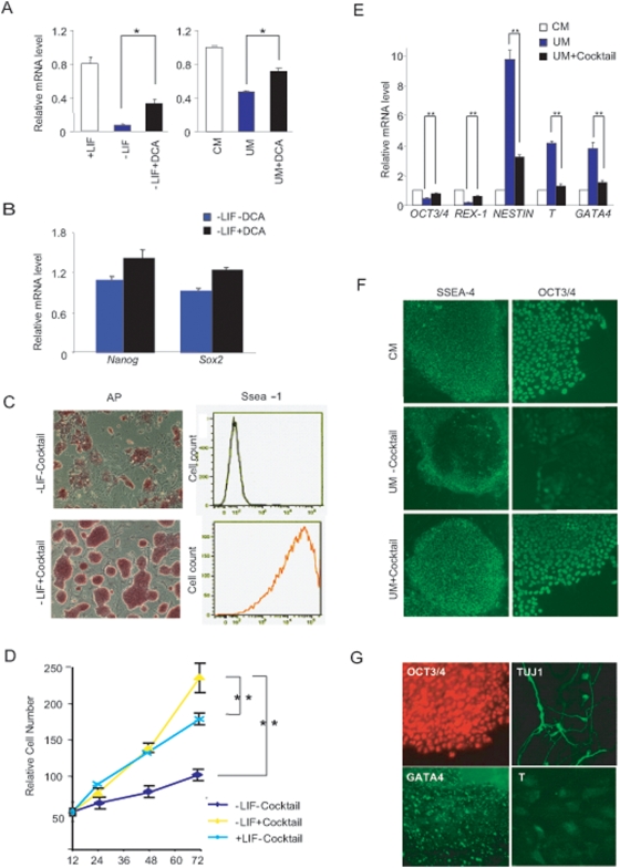
Recent studies have suggested distinctive biological properties and signaling mechanisms between human and mouse embryonic stem cells (hESCs and mESCs). Herein we report that Shp2, a protein tyrosine phosphatase with two SH2 domains, has a conserved role in orchestration of intracellular signaling cascades resulting in initiation of differentiation in both hESCs and mESCs. Homozygous deletion of Shp2 in mESCs inhibited differentiation into all three germ layers, and siRNA-mediated knockdown of Shp2 expression in hESCs led to a similar phenotype of impaired differentiation. A small molecule inhibitor of Shp2 enzyme suppressed both hESC and mESC differentiation capacity. Shp2 modulates Erk, Stat3 and Smad pathways in ES cells and, in particular, Shp2 regulates BMP4-Smad pathway bi-directionally in mESCs and hESCs. These results reveal a common signaling mechanism shared by human and mouse ESCs via Shp2 modulation of overlapping and divergent pathways.


Conflict of interest statement

Competing Interests: The authors have declared that no competing interests exist.

Figures

Figure 1
Figure 1. Establishment of Shp2−/− mESC lines.
(A) Genotyping of genomic DNA (upper panel). The wild-type (wt) and exon 4Δ alleles were detected by PCR. Immunoblotting analysis for Shp2 confirmed the PCR results (lower panel), indicating that clones # 3, 7 and 11 are homozygous (Shp2−/−) mutants. (B) Alkaline phosphatase (AP) staining of wt and Shp2−/− mESCs cultured on feeder cells. Scale bars, 50 µm. (C) FACS analysis of Ssea-1 expression on mESCs in feeder-free culture.
Figure 2
Figure 2. Shp2−/− mES cells exhibit reduced differentiation capacity with improved self-renewal potential.
(A) Immunostaining for Nanog, Oct3/4, Sox2, and flow cytometry for Ssea-1 in primary (1st) EBs cultured in LIF-free suspension medium for 8 days. Scale bars, 10 µm. (B) 1st EBs were dissociated into single cells and re-seeded at the density of 1×10⁶ cells/ml onto feeder cells and cultured in ES cell standard medium (+LIF) for colony forming assay. The cells were fixed and AP-stained. The AP-positive cells were counted under a bright field microscope. Scale bars, 200 µm. (C) 1st EBs were dissociated into single cells and re-seeded at the density of 1×10⁶ cells/ml in the same medium as 1st EBs for formation of secondary (2nd) EBs. Scale bars, 200 µm. The number of 2nd EBs was counted under a bright field microscope. (D) Total RNA was extracted from mESCs (day 0) and differentiating EBs at day10, and qRT-PCR was performed, and the relative values were normalized by CPH. Relative value changes of Shp2−/− over wt mESCs were indicated (n>3, * P<0.05, ** P<0.01).
Figure 3
Figure 3. Shp2 knockdown in hESCs leads to impaired differentiation.
(A) Immunoblotting and qRT-PCR indicate efficient Shp2 knockdown by 80% in hESCs. H14 cells were transfected with Shp2-specific (+siShp2) or non-specific siRNA (−siShp2), and cells were cultured on matrigel-coated dishes in CM for 2 days, total cell lysates were immunoblotted with the indicated antibodies or total RNAs were extracted for qRT-PCR (n = 3, ** P<0.01). (B) Immunostaining of NANOG, OCT3/4, SOX2 in hESCs. After transfection, H14 cells were cultured in CM for 2 days, then the medium were changed to UM to induce differentiation for additional 6 days. Scale bars, 10 µm. (C) Total RNAs were extracted from hESCs cultured in CM or UM, qRT-PCR was performed, and the relative values of Shp2 knockdown over control hESCs were indicated (n = 3, * P<0.05, ** P<0.01).
Figure 4
Figure 4. Shp2 modulates various signaling pathways in mES and hES cells.
(A) LIF/BMP4 signaling in mESCs. mESCs were stimulated by LIF (1,000 units/ml) or BMP4 (25 ng/ml) for 15 mins after overnight starvation. Total cell lysates were immunoblotted with the indicated antibodies. (B) bFGF/BMP4 signaling in hESCs (H14). The indicated antibodies were used to analyze proteins in hESCs stimulated by bFGF (50 ng/ml, 15 mins) or BMP4 (25 ng/ml, 30 min) after 6-hour starvation. (C, D) mESCs (left) or hESCs (right) were treated with BMP4 for 60 mins and total RNAs were analyzed for Id expression levels (Mean±SEM, n = 3). (a, c, e) mESCs; (b, d, f) hESCs.
Figure 5
Figure 5. Chemical Inhibitors suppress mESC and hESC differentiation.
(A) DCA partially inhibits differentiation in both mESCs (left panel) and hESCs (right panel) monitored by qRT-PCR analysis of Oct3/4 expression. (B) qRT-PCR analysis of pluripotent cell markers Nanog and Sox2. mESCs were incubated in feeder-free and LIF-free medium in the absence or presence of 50 µM DCA for 6 days and total RNAs were extracted for analysis (Mean±SEM, n = 3). (C) AP staining (left panel) and Ssea-1 flow cytometry (right panel) of mESCs treated with or without cocktail inhibitors. Scale bars, 50 µm. (D) mESCs cultured in feeder-free and LIF-free medium with cocktail inhibitors exhibited comparable proliferation rate as cells cultured in standard mESC medium (+LIF). (E) QRT-PCR analysis of pluripotent cell markers OCT3/4 and REX-1, or differentiated cell markers NESTIN, T and GATA4. hESCs were incubated in CM, or UM in the absence or presence of cocktail inhibitors as indicated (25 µM DCA, 25 µM PD and 0.5 µM Bio) for 6 days (Mean±SEM, n = 3). (F) Morphological analysis of hESCs performed by immunostaining of OCT3/4, and SSEA-4. Scale bar, 10 µm. (G) Removal of cocktail inhibitors restored differentiation capacity. hESC cells maintained with the cocktail inhibitors were incubated in UM without inhibitors for 8 days and allowed for differentiation under standard procedures. Cells were immunostained for TUJ1+ neurons, GATA4+ endoderm cell lineage or T+ mesoderm cell lineage, and hES cells incubated in CM were immunostained for OCT3/4. Scale bar, 5 µm.

References

    1. Thomson JA, Itskovitz-Eldor J, Shapiro SS, Waknitz MA, Swiergiel JJ, et al. Embryonic stem cell lines derived from human blastocysts. Science. 1998;282:1145–1147. - PubMed
    1. Boyer LA, Lee TI, Cole MF, Johnstone SE, Levine SS, et al. Core transcriptional regulatory circuitry in human embryonic stem cells. Cell. 2005;122:947–956. - PMC - PubMed
    1. Loh YH, Wu Q, Chew JL, Vega VB, Zhang W, et al. The Oct4 and Nanog transcription network regulates pluripotency in mouse embryonic stem cells. Nat Genet. 2006;38:431–440. - PubMed
    1. Lee TI, Jenner RG, Boyer LA, Guenther MG, Levine SS, et al. Control of developmental regulators by Polycomb in human embryonic stem cells. Cell. 2006;125:301–313. - PMC - PubMed
    1. Sato N, Sanjuan IM, Heke M, Uchida M, Naef F, et al. Molecular signature of human embryonic stem cells and its comparison with the mouse. Dev Biol. 2003;260:404–413. - PubMed

Publication types

MeSH terms

Substances